J Korean Soc Radiol.
2010 Oct;63(4):307-310.
A Cerebral Air Embolism That Developed Following Defecation in a Patient with Extensive Pulmonary Tuberculosis: A Case Report
- Affiliations
-
- 1Department of Radiology, College of Medicine, Hanyang University, Korea. dwpark@hanyang.ac.kr
Abstract
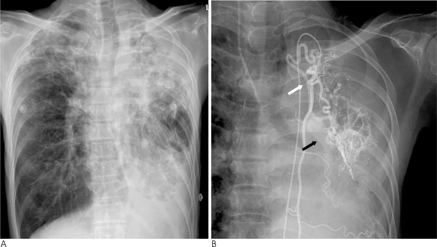
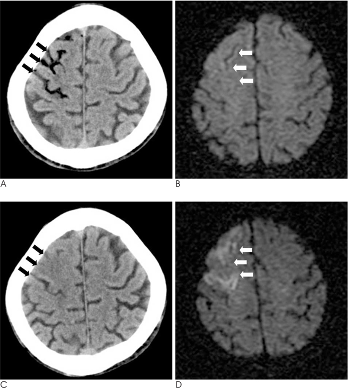
- Cerebral air embolisms generally result from invasive procedures such as a percutaneous needle biopsy, chest tube insertion, central venous catheter access or removal, operations and so on. Likewise, they are mostly iatrogenically induced and present various degrees of severity depending on the number of air bubbles. With the exception of divers, the occurrence of a cerebral air embolism in the absence of invasive procedures is very rare. We report a case of a cerebral air embolism that developed following defecation and was detected by CT in a patient with extensive pulmonary tuberculosis.
MeSH Terms
Figure
Reference
-
1. Kau T, Rabitsch E, Celedin S, Habernig SM, Weber JR, Hausegger KA. When coughing can cause stroke-a case-based update on cerebral air embolism complicating biopsy of the lung. Cardiovasc Intervent Radiol. 2008; 31:848–853.2. Yamashita Y, Mukaida H, Hirabayashi N, Takiyama W. Cerebral air embolism after intrathoracic anti-cancer drug administration. Ann Thorac Surg. 2006; 82:1121–1123.3. Hsi DH, Thompson TN, Fruchter A, Collins MS, Lieberg OU, Boepple H. Simultaneous coronary and cerebral air embolism after CT-guided core needle biopsy of the lung. Tex Heart Inst J. 2008; 35:472–474.4. Muth CM, Shank ES. Gas embolism. N Engl J Med. 2000; 342:476–482.5. Helps SC, Parsons DW, Reilly PL, Gorman DF. The effect of gas emboli on rabbit cerebral blood flow. Stroke. 1990; 21:94–99.6. Murphy BP, Harford FJ, Cramer FS. Cerebral air embolism resulting from invasive medical procedures. Treatment with hyperbaric oxygen. Ann Surg. 1985; 201:242–245.7. Ashizawa K. Possible airflow around the needle in lung biopsy. AJR Am J Roentgenol. 2005; 185:553.
- Full Text Links
- Actions
-
Cited
- CITED
-
- Close
- Share
- Similar articles
-
- Cerebral Air Embolism in a Patient with a Tuberculous-Destroyed Lung during Commercial Air Travel: A Case Report
- Cerebral Air Embolism Associated with Pulmonary Tuberculosis
- Cerebral Air Embolism Following a Gastroscopy
- Cerebral Air Embolism and Cardiomyopathy Secondary to Large Bulla Rupture during a Pulmonary Function Test
- Stroke Caused by Cerebral Air Embolism after Central Venous Catheter Removal: A Case Report


