J Korean Acad Prosthodont.
2016 Jul;54(3):298-305. 10.4047/jkap.2016.54.3.298.
Implant esthetic restoration with bone graft in the extended maxillary anterior area: A case report
- Affiliations
-
- 1Major in Dentistry, Department of Medical Science, Hanyang University, Seoul, Republic of Korea. leeys@hanyang.ac.kr
- KMID: 2344871
- DOI: http://doi.org/10.4047/jkap.2016.54.3.298
Abstract
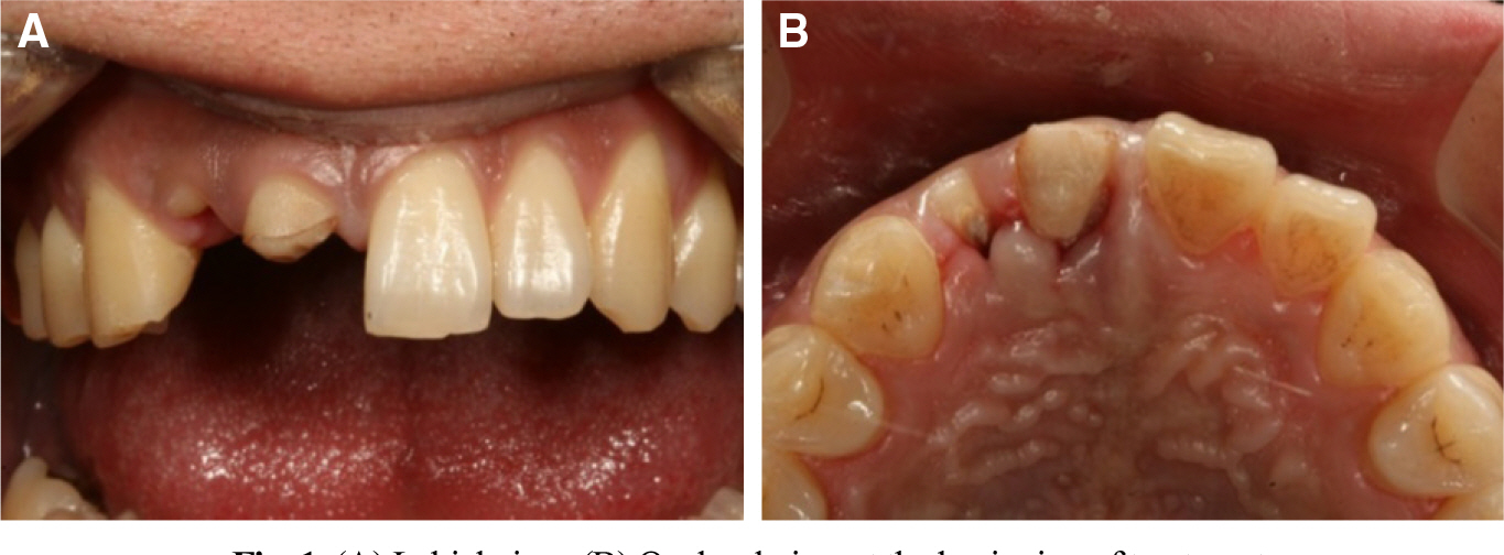
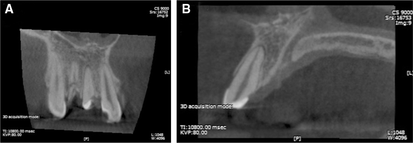
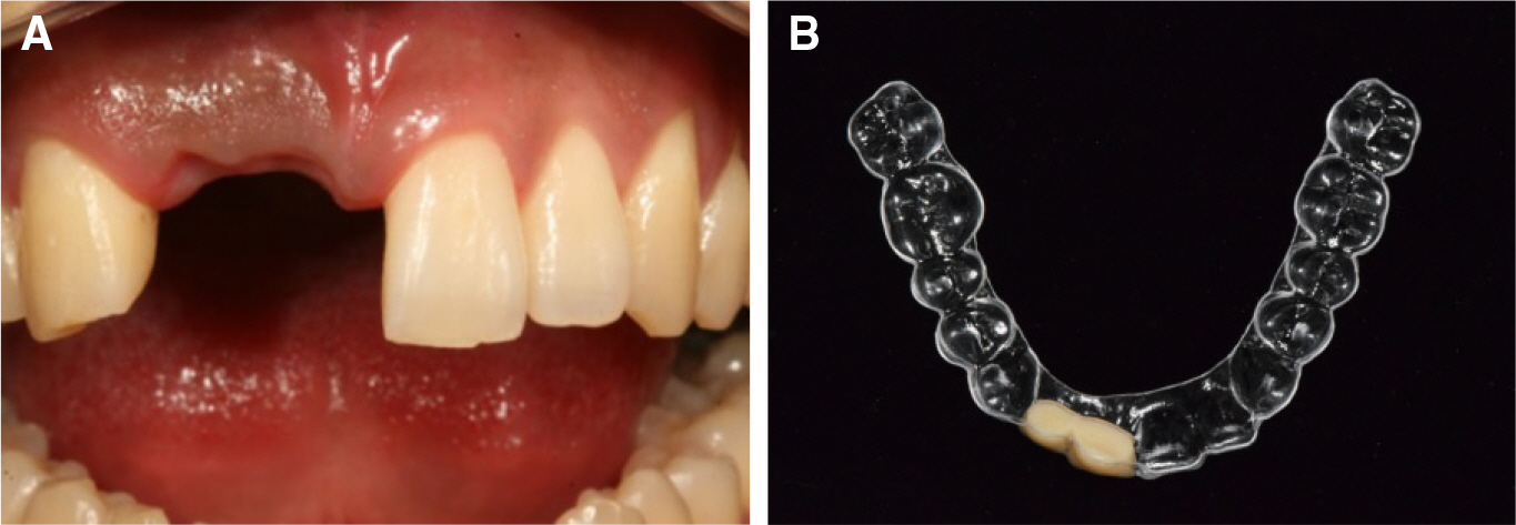
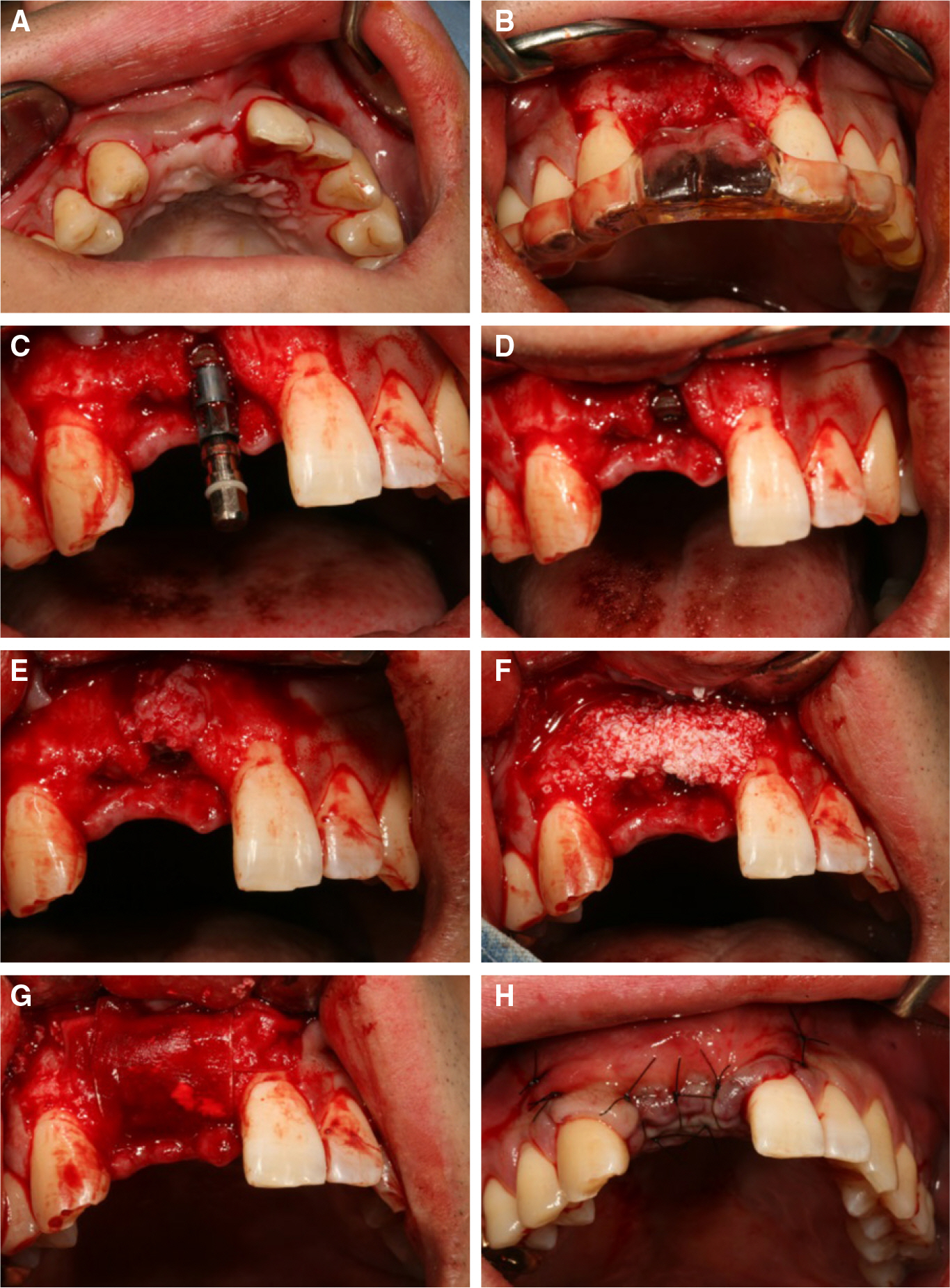
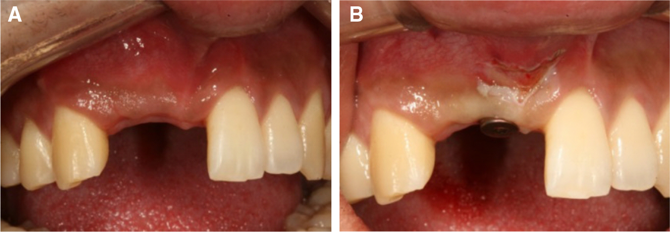
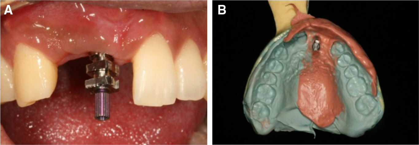
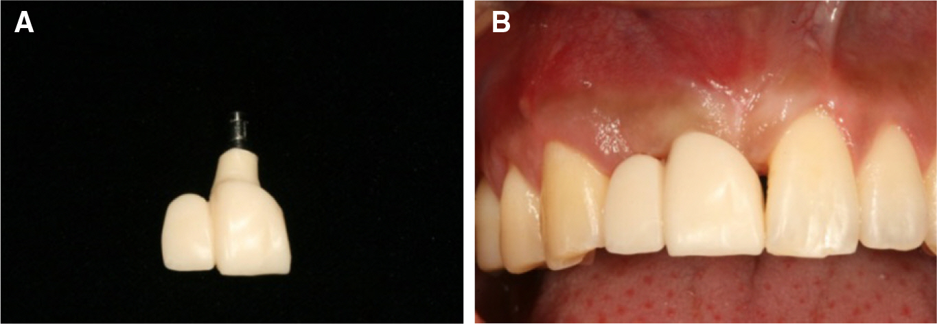
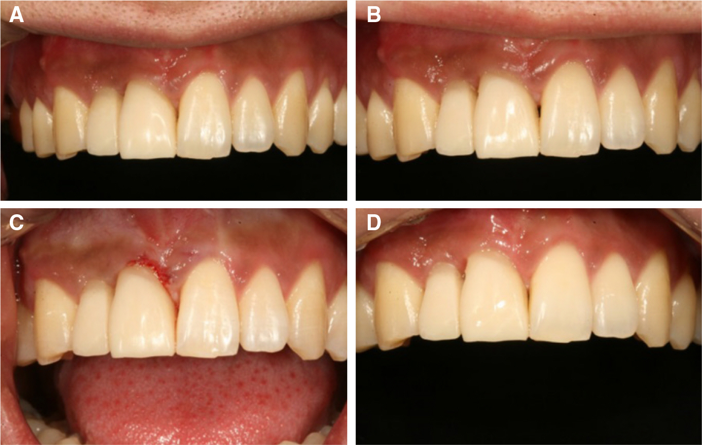
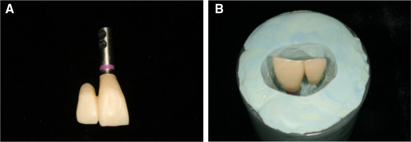
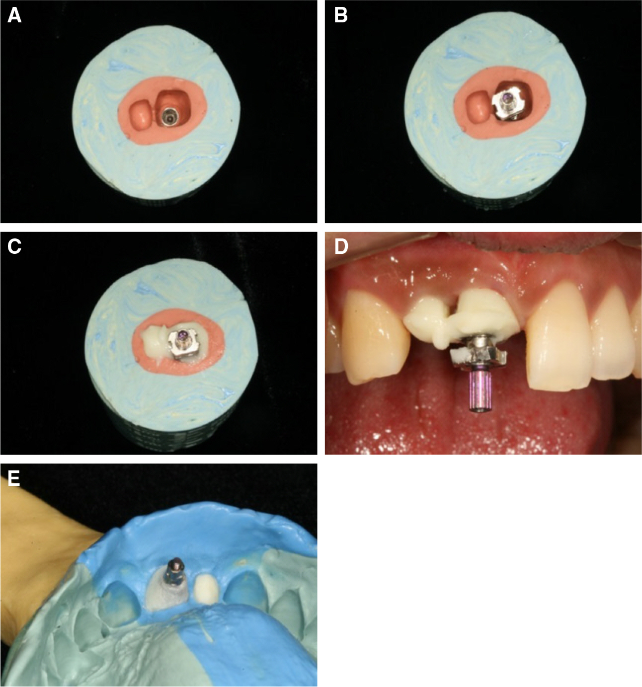
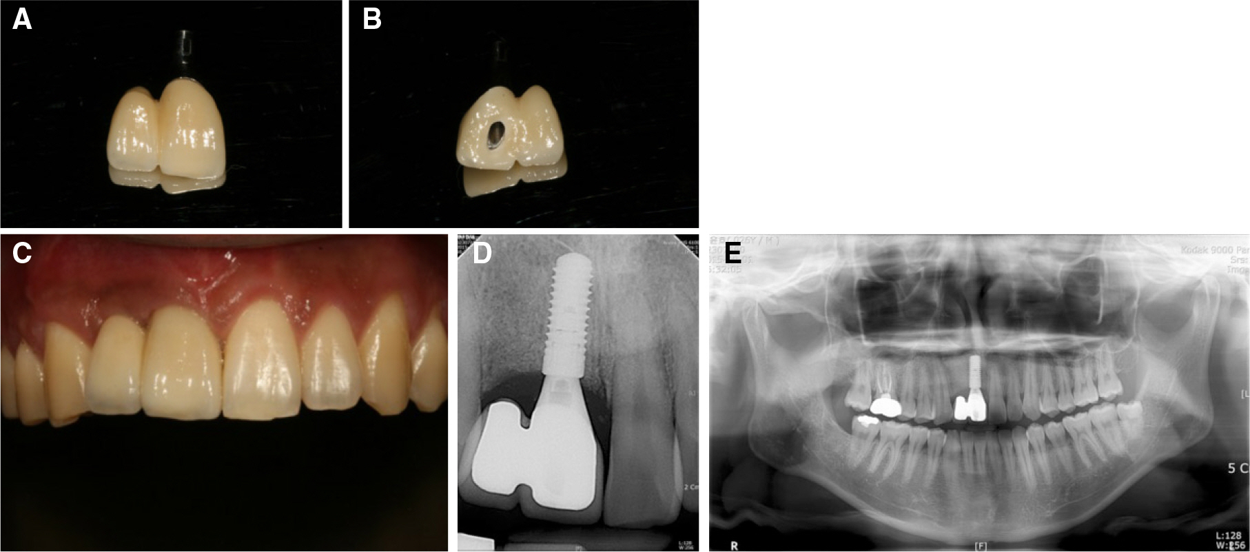
- The maxillary anteriors play an important role in esthetics. Therefore after extraction, it is crucial to preserve the hard tissue and soft tissue in order to promote esthetics of restoration. There are several challenges when restoring the maxillary anteriors via implant. Some of the challenges are be maintaining consistency with neighboring teeth in terms of shade, form, and texture : as well as having harmonious emergency with the gingival margin. In this case, a traumatized patient with crown-root fracture of the maxillary central and lateral incisors is presented. The cracked teeth were extracted, and implants were inserted with bone grafts to compensate the volume of damaged area of the maxillary anterior. Cantilever implant prosthetics were planned while precise adjustments to the gingival area were made using customized impression coping to perform the esthetic restorations. The final outcome of the treatment was satisfying in both esthetic and utilitarian perspective.
Figure
Reference
-
1.Santosa RE. Provisional restoration options in implant dentistry. Aust Dent J. 2007. 52:234–42.2.Buser D., Chen ST., Weber HP., Belser UC. Early implant placement following single-tooth extraction in the esthetic zone: biologic rationale and surgical procedures. Int J Periodontics Restorative Dent. 2008. 28:441–51.3.Buser D., Martin W., Belser UC. Optimizing esthetics for implant restorations in the anterior maxilla: anatomic and surgical considerations. Int J Oral Maxillofac Implants. 2004. 19:43–61.4.Dawson T., Chen ST. The SAC classification in implant dentistry. Berlin: Quintessence Publishing;2009.5.Tarnow DP., Cho SC., Wallace SS. The effect of inter-implant distance on the height of inter-implant bone crest. J Periodontol. 2000. 71:546–9.
Article6.Kan JY., Rungcharassaeng K. Interimplant papilla preservation in the esthetic zone: a report of six consecutive cases. Int J Periodontics Restorative Dent. 2003. 23:249–59.7.Daoudi MF., Setchell DJ., Searson LJ. A laboratory investigation of the repositioning impression coping technique at the implant level for single-tooth implants. Eur J Prosthodont Restor Dent. 2003. 11:23–8.8.Buser D., Halbritter S., Hart C., Bornstein MM., Grütter L., Chappuis V., Belser UC. Early implant placement with simultaneous guided bone regeneration following single-tooth extraction in the esthetic zone: 12-month results of a prospective study with 20 consecutive patients. J Periodontol. 2009. 80:152–62.
Article9.Buser D., Belser UC. Correct three-dimensional implant placement: the concept of danger and comfort zones. Quintessence Publishing;2008.10.Jensen SS., Broggini N., Hjørting-Hansen E., Schenk R., Buser D. Bone healing and graft resorption of autograft, anorganic bovine bone and beta-tricalcium phosphate. A histologic and histomorphometric study in the mandibles of minipigs. Clin Oral Implants Res. 2006. 17:237–43.
Article
- Full Text Links
- Actions
-
Cited
- CITED
-
- Close
- Share
- Similar articles
-
- Esthetic restoration in continuous maxillary anterior area using immediate implant placement: A case report
- Implant Surgery with Both Sinus Bone Graft in the Maxillary edentulous patient: A Case Report
- Decoronation and implant restoration of ankylosed tooth resulted from anterior avulsion: A case report
- Outcome Evaluation of an Immediately Placed Maxillary Anterior Single-Tooth Implant Using Objective Esthetic Criteria: Case Report
- Esthetic implant restoration in the maxillary anterior missing area with palatal defect of the alveolar bone: a case report

