J Korean Acad Prosthodont.
2016 Jul;54(3):210-217. 10.4047/jkap.2016.54.3.210.
Evaluation of marginal and internal gap under model-free monolithic zirconia restoration fabricated by digital intraoral scanner
- Affiliations
-
- 1Department of Prosthodontics, Ewha Womans University Mokdong Hospital, Seoul, Republic of Korea.
- 2Department of Prosthodontics and Dental Research Institute, Seoul National University Gwanak Dental Hospital, Seoul, Republic of Korea. jimarn@gmail.com
- KMID: 2344858
- DOI: http://doi.org/10.4047/jkap.2016.54.3.210
Abstract
- PURPOSE
The aim of this study was to evaluate the marginal and internal adaptation of monolithic zirconia restoration made without physical model by digital intraoral scanner.
MATERIALS AND METHODS
A prospective clinical trial was performed on 11 restorations as a pilot study. The monolithic zirconia restorations were fabricated after digital intraoral impression taking by intraoral scanner (TRIOS, 3shape, Copenhagen, Denmark), computer-aided designing, and milling manufacturing process. Completed zirconia crowns were tried in the patients' mouth and a replica technique was used to acquire the crown-abutment replica. The absolute marginal discrepancy, marginal gap, and internal gap of axial, line angle, and occlusal part were measured after sectioning the replica in the mesiodistal and buccolingual direction. Statistical analysis was performed using Kruskal-Wallis and Mann-Whitney U test (α=.05).
RESULTS
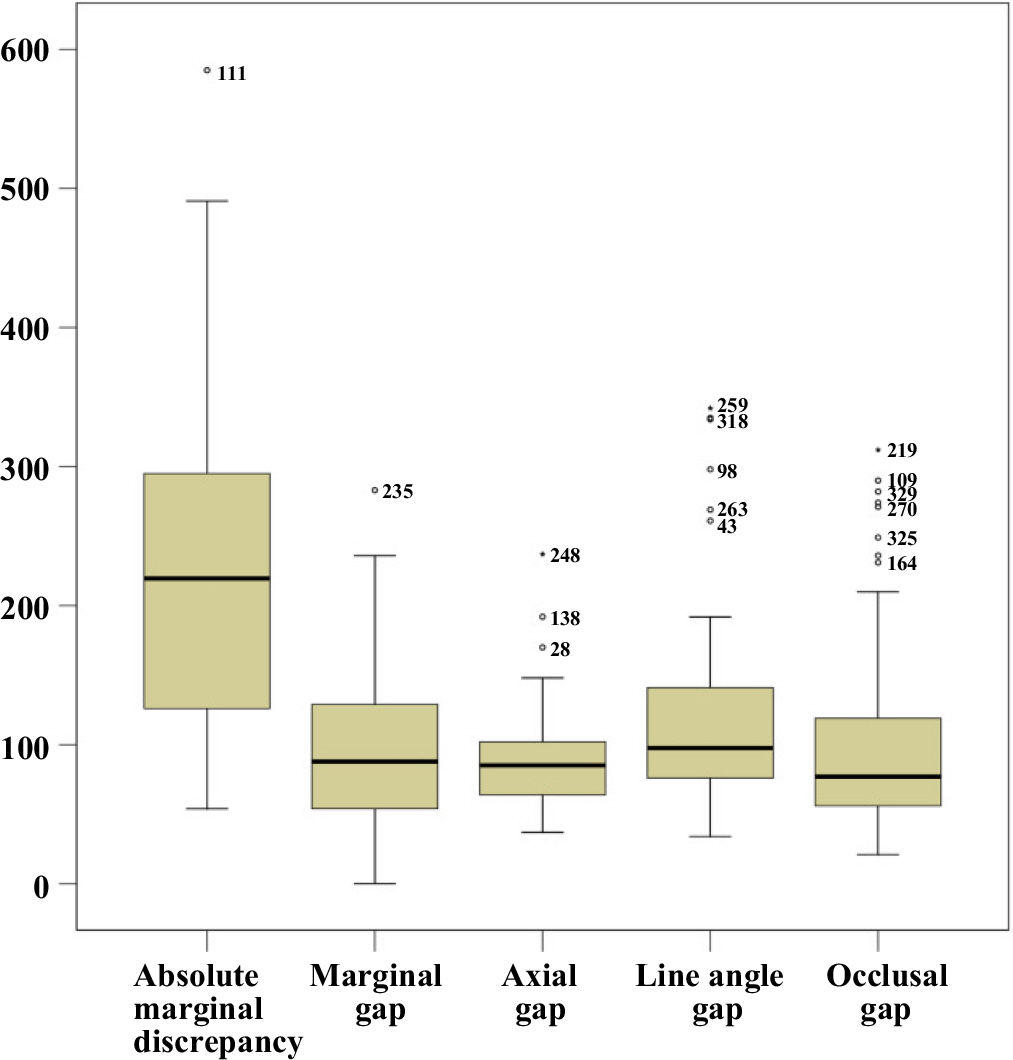
From the adaptation analysis by replica, the statistically significant difference was not found between mesiodistal and buccolingual sections (P>.05), but there was significant difference among the measurement location (P<.01). The amount of absolute marginal discrepancy was larger than those of marginal gap and internal gap (P<.01).
CONCLUSION
Within the limitations of this study, the adaptation accuracy of model-free monolithic zirconia restoration fabricated by intraoral scanner exhibited clinically acceptable result. However, the margin of zirconia crown showed tendency of overcontour and cautious clinical application and follow up is necessary.
Keyword
MeSH Terms
Figure
Reference
-
1.Vagkopoulou T., Koutayas SO., Koidis P., Strub JR. Zirconia in dentistry: Part 1. Discovering the nature of an upcoming bioceramic. Eur J Esthet Dent. 2009. 4:130–51.2.Piconi C., Maccauro G. Zirconia as a ceramic biomaterial. Biomaterials. 1999. 20:1–25.
Article3.Covacci V., Bruzzese N., Maccauro G., Andreassi C., Ricci GA., Piconi C., Marmo E., Burger W., Cittadini A. In vitro evaluation of the mutagenic and carcinogenic power of high purity zirconia ceramic. Biomaterials. 1999. 20:371–6.
Article4.Koutayas SO., Vagkopoulou T., Pelekanos S., Koidis P., Strub JR. Zirconia in dentistry: part 2. Evidence-based clinical breakthrough. Eur J Esthet Dent. 2009. 4:348–80.5.Raigrodski AJ., Chiche GJ., Potiket N., Hochstedler JL., Mohamed SE., Billiot S., Mercante DE. The efficacy of posterior three-unit zirconium-oxide-based ceramic fixed partial dental prostheses: a prospective clinical pilot study. J Prosthet Dent. 2006. 96:237–44.
Article6.Roediger M., Gersdorff N., Huels A., Rinke S. Prospective evaluation of zirconia posterior fixed partial dentures: four-year clinical results. Int J Prosthodont. 2010. 23:141–8.7.Rinke S., Gersdorff N., Lange K., Roediger M. Prospective evaluation of zirconia posterior fixed partial dentures: 7-year clinical results. Int J Prosthodont. 2013. 26:164–71.
Article8.Sailer I., Gottnerb J., Kanelb S., Hammerle CH. Randomized controlled clinical trial of zirconia-ceramic and metal-ceramic posterior fixed dental prostheses: a 3-year follow-up. Int J Prosthodont. 2009. 22:553–60.9.Albashaireh ZS., Ghazal M., Kern M. Two-body wear of different ceramic materials opposed to zirconia ceramic. J Prosthet Dent. 2010. 104:105–13.
Article10.Jung YS., Lee JW., Choi YJ., Ahn JS., Shin SW., Huh JB. A study on the in-vitro wear of the natural tooth structure by opposing zirconia or dental porcelain. J Adv Prosthodont. 2010. 2:111–5.11.Suttor D., Bunke K., Hoescheler S., Hauptmann H., Hertlein G. LA-VA—the system for all-ceramic ZrO2 crown and bridge frameworks. Int J Comput Dent. 2001. 4:195–206.12.Filser F., Kocher P., Weibel F., Lüthy H., Schärer P., Gauckler LJ. Reliability and strength of all-ceramic dental restorations fabricated by direct ceramic machining (DCM). Int J Comput Dent. 2001. 4:89–106.13.Ender A., Mehl A. Full arch scans: conventional versus digital impressions-an in-vitro study. Int J Comput Dent. 2011. 14:11–21.14.Seelbach P., Brueckel C., Wöstmann B. Accuracy of digital and conventional impression techniques and workflow. Clin Oral Investig. 2013. 17:1759–64.
Article15.Reich S., Wichmann M., Nkenke E., Proeschel P. Clinical fit of all-ceramic three-unit fixed partial dentures, generated with three different CAD/CAM systems. Eur J Oral Sci. 2005. 113:174–9.
Article16.Laurent M., Scheer P., Dejou J., Laborde G. Clinical evaluation of the marginal fit of cast crowns-validation of the silicone replica method. J Oral Rehabil. 2008. 35:116–22.17.Rahme HY., Tehini GE., Adib SM., Ardo AS., Rifai KT. In vitro evaluation of the "replica technique" in the measurement of the fit of Procera crowns. J Contemp Dent Pract. 2008. 9:25–32.18.Kohorst P., Brinkmann H., Dittmer MP., Borchers L., Stiesch M. Influence of the veneering process on the marginal fit of zirconia fixed dental prostheses. J Oral Rehabil. 2010. 37:283–91.
Article19.Reich S., Uhlen S., Gozdowski S., Lohbauer U. Measurement of cement thickness under lithium disilicate crowns using an impression material technique. Clin Oral Investig. 2011. 15:521–6.
Article20.Syrek A., Reich G., Ranftl D., Klein C., Cerny B., Brodesser J. Clinical evaluation of all-ceramic crowns fabricated from intraoral digital impressions based on the principle of active wavefront sampling. J Dent. 2010. 38:553–9.
Article21.Holmes JR., Bayne SC., Holland GA., Sulik WD. Considerations in measurement of marginal fit. J Prosthet Dent. 1989. 62:405–8.
Article22.McLean JW., von Fraunhofer JA. The estimation of cement film thickness by an in vivo technique. Br Dent J. 1971. 131:107–11.
Article23.Schaerer P., Sato T., Wohlwend A. A comparison of the marginal fit of three cast ceramic crown systems. J Prosthet Dent. 1988. 59:534–42.
Article24.Pera P., Gilodi S., Bassi F., Carossa S. In vitro marginal adaptation of alumina porcelain ceramic crowns. J Prosthet Dent. 1994. 72:585–90.
Article25.Groten M., Girthofer S., Pröbster L. Marginal fit consistency of copy-milled all-ceramic crowns during fabrication by light and scanning electron microscopic analysis in vitro. J Oral Rehabil. 1997. 24:871–81.
Article26.Sulaiman F., Chai J., Jameson LM., Wozniak WT. A comparison of the marginal fit of In-Ceram, IPS Empress, and Procera crowns. Int J Prosthodont. 1997. 10:478–84.27.Boening KW., Wolf BH., Schmidt AE., Kästner K., Walter MH. Clinical fit of Procera AllCeram crowns. J Prosthet Dent. 2000. 84:419–24.
Article28.Sua ′rez MJ., Gonza ′lez de Villaumbrosia P., Pradl′es G., Lozano JF. Comparison of the marginal fit of Procera AllCeram crowns with two finish lines. Int J Prosthodont. 2003. 16:229–32.29.Yeo IS., Yang JH., Lee JB. In vitro marginal fit of three all-ceramic crown systems. J Prosthet Dent. 2003. 90:459–64.
Article30.Bindl A., Mörmann WH. Marginal and internal fit of all-ceramic CAD/CAM crown-copings on chamfer preparations. J Oral Rehabil. 2005. 32:441–7.
Article31.Nakamura T., Tanaka H., Kinuta S., Akao T., Okamoto K., Wakabayashi K., Yatani H. In vitro study on marginal and internal fit of CAD/CAM all-ceramic crowns. Dent Mater J. 2005. 24:456–9.
Article32.Brawek PK., Wolfart S., Endres L., Kirsten A., Reich S. The clinical accuracy of single crowns exclusively fabricated by digital workflow—the comparison of two systems. Clin Oral Investig. 2013. 17:2119–25.
Article33.Park JM., Hong YS., Park EJ., Heo SJ., Oh N. Clinical evaluations of cast gold alloy, machinable zirconia, and semiprecious alloy crowns: A multicenter study. J Prosthet Dent. 2016. 115:684–91.
Article34.Kohorst P., Brinkmann H., Li J., Borchers L., Stiesch M. Marginal accuracy of four-unit zirconia fixed dental prostheses fabricated using different computer-aided design/computer-aided manufacturing systems. Eur J Oral Sci. 2009. 117:319–25.
Article
- Full Text Links
- Actions
-
Cited
- CITED
-
- Close
- Share
- Similar articles
-
- In-vitro evaluation of marginal and internal fit of 3-unit monolithic zirconia restorations fabricated using digital scanning technologies
- Assessment of the fit of zirconia-based prostheses fabricated with two different scan methods
- Superimposition: a simple method to minimize occlusal adjustment of monolithic restoration
- Comparison of marginal and internal fit of 3-unit monolithic zirconia fixed partial dentures fabricated from solid working casts and working casts from a removable die system
- Immediate loading of mandibular single implant by using surgical guide and modeless digital prosthesis: a case report


