Ann Dermatol.
2016 Aug;28(4):509-510. 10.5021/ad.2016.28.4.509.
Scar Sarcoidosis Induced by Pulsed Dye Laser Treatment
- Affiliations
-
- 1Department of Dermatology, Chungnam National University School of Medicine, Daejeon, Korea. Jhoon@cnu.ac.kr
- KMID: 2344829
- DOI: http://doi.org/10.5021/ad.2016.28.4.509
Abstract
- No abstract available.
MeSH Terms
Figure
-
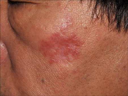
Fig. 1 Erythematous plaque on the left cheek with a white atrophic area in the lower part of the lesion as the trace of an old scar.
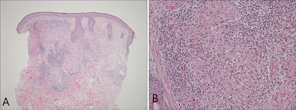
Fig. 2 (A) Well-demarcated multiple islands of epithelioid histiocytes, which are surrounded by fairly dense lymphocytic infiltrate (H&E, ×40). (B) High-power view showing typical sarcoidal granuloma with mononuclear cell infiltrates (H&E, ×200).
Reference
-
1. Haimovic A, Sanchez M, Judson MA, Prystowsky S. Sarcoidosis: a comprehensive review and update for the dermatologist: part I. Cutaneous disease. J Am Acad Dermatol. 2012; 66:699.e1–699.e18.2. Sorabjee JS, Garje R. Reactivation of old scars: inevitably sarcoid. Postgrad Med J. 2005; 81:60–61.
Article3. Erceg A, de Jong EM, van de Kerkhof PC, Seyger MM. The efficacy of pulsed dye laser treatment for inflammatory skin diseases: a systematic review. J Am Acad Dermatol. 2013; 69:609–615.e8.
Article4. Emer J, Uslu U, Waldorf H. Improvement in lupus pernio with the successive use of pulsed dye laser and nonablative fractional resurfacing. Dermatol Surg. 2014; 40:201–202.
Article5. Green JJ, Lawrence N, Heymann WR. Generalized ulcerative sarcoidosis induced by therapy with the flashlamp-pumped pulsed dye laser. Arch Dermatol. 2001; 137:507–508.
- Full Text Links
-
- Actions
-
Cited
- CITED
-
- Close
- Share
-
- Similar articles
-
- Facial Scar Treatment Using 585nm Pulsed Dye Laser
- 595nm Pulsed Dye Laser (Vbeam(R)) Treatment of Hypertrophic Scar
- Treatment of Striae Distensae by Thermage and 585-nm Pulsed Dye Laser(V-star(R))
- Treatment of Verrucae with Flashlamp-pumped Pulsed Dye Laser
- Combination Therapy with Intralesional Interferon α-2b and Pulsed Dye Laser for the Treatment of Periungual Warts
