Synchronous Occurrence of Primary Cutaneous Anaplastic Large Cell Lymphoma and Squamous Cell Carcinoma
- Affiliations
-
- 1Department of Dermatology, Samsung Medical Center, Sungkyunkwan University School of Medicine, Seoul, Korea. dylee@skku.edu
- KMID: 2344821
- DOI: http://doi.org/10.5021/ad.2016.28.4.491
Abstract
- CD30+ lymphoproliferative disorders (LPD) represent a spectrum of T-cell lymphoma including lymphomatoid papulosis and anaplastic large cell lymphoma (ALCL). Epidermis overlying cutaneous CD30+ LPD often shows epidermal hyperplasia, hyperkeratosis, crusting, and ulceration and it is difficult to distinguish from carcinoma such as keratoacanthoma (KA) or squamous cell carcinoma (SCC). Several cases of pseudocarcinomatous hyperplasia mimicking KA or SCC in CD30+ LPD have been reported. The relationship between CD30+ LPD and epithelial proliferations has not yet well understood. It was reported that a variety of mediators, including epidermal growth factor (EGF), transforming growth factor-α and EGFR from CD30+ LPD could attribute to epidermal hyperplasia. However, separate and distinct SCC occurring in CD30+ LPD has rarely been reported. Herein, we present a rare case of coexistence of SCC and cutaneous ALCL located on the same region.
Keyword
MeSH Terms
Figure
-
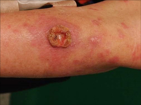
Fig. 1 A 15 mm diameter ulcerative tumor with erythematous plaques and papules on the left forearm.

Fig. 2 (A) Histological analysis of the shave biopsy specimen shows exo-endophytic epidermal proliferation with hyperkeratosis, horn cysts, and microabscesses and dense cellular infiltrates in dermis (H&E, ×40). (B) Invasive atypical cells dissecting collagen bundle suggested squamous cell carcinoma (arrows; H&E, ×200). (C) Large atypical cells were obscured by a massive infiltrate of neutrophils and eosinophils in dermis (arrows; H&E, ×200). (D) Large atypical cells are positive for CD30 (CD30, ×200). (E) Expression of Ki-67 was diffuse in epidermis and large cells of dermis. (F) The expression of p53 was diffuse and scattered throughout epidermis.
Reference
-
1. Cespedes YP, Rockley PF, Flores F, Ruiz P, Kaiser MR, Elgart GW. Is there a special relationship between CD30-positive lymphoproliferative disorders and epidermal proliferation? J Cutan Pathol. 2000; 27:271–275.
Article2. Kawachi Y, Taguchi S, Fujisawa Y, Furuta J, Nakamura Y, Ishii Y, et al. Epidermal pseudocarcinomatous hyperplasia with underlying epidermal growth factor-producing cutaneous CD30-positive lymphoproliferative disorder. J Eur Acad Dermatol Venereol. 2009; 23:181–183.
Article3. Yanagi T, Shimizu T, Kodama K, Nemoto-Hasebe I, Kasai M, Shimizu H. CD30-positive primary cutaneous anaplastic large-cell lymphoma and definite squamous cell carcinoma. Clin Exp Dermatol. 2009; 34:e293–e294.
Article4. Price A, Miller JH, Junkins-Hopkins JM. Pseudocarcinomatous hyperplasia in anaplastic large cell lymphoma, a mimicker of poorly differentiated squamous cell carcinoma: report of a case and review of the literature. J Cutan Pathol. 2015; 42:863–869.
Article5. Courville P, Wechsler J, Thomine E, Vergier B, Fonck Y, Souteyrand P, et al. Pseudoepitheliomatous hyperplasia in cutaneous T-cell lymphoma. A clinical, histopathological and immunohistochemical study with particular interest in epithelial growth factor expression. The French Study Group on Cutaneous Lymphoma. Br J Dermatol. 1999; 140:421–426.
Article6. Cowley GP, Smith JA, Gusterson BA. Increased EGF receptors on human squamous carcinoma cell lines. Br J Cancer. 1986; 53:223–229.
Article7. Fernandez-Flores A. CD30+ cell population in common keratoacanthomas: a study of 21 cases. Rom J Morphol Embryol. 2008; 49:159–162.8. Fernandez-Flores A. CD30+ cells in regressing keratoacanthoma and in non-keratoacanthomatous squamous cell carcinoma. Bratisl Lek Listy. 2008; 109:508–512.
- Full Text Links
-
- Actions
-
Cited
- CITED
-
- Close
- Share
-
- Similar articles
-
- Dermatofibroma in Patient with Relapsing Primary Cutaneous Anaplastic Large Cell Lymphoma
- A Case of Primary Cutaneous Anaplastic Large Cell Lymphoma on the Dorsum of the Hand
- Primary Cutaneous CD30+ Anaplastic Large Cell Lymphoma That Developed after Lymphomatoid Papulosis
- A Case of Multifocal Primary Cutaneous Anaplastic Large Cell Lymphoma Managed without Surgical Treatment
- A Case of Primary Cutaneous CD30+ Anaplastic Large Cell Lymphoma Presenting with Numerous Patches, Papules, and Nodules
