Korean Circ J.
2016 Jul;46(4):530-535. 10.4070/kcj.2016.46.4.530.
Atrial Electromechanical Coupling in Patients with Lichen Planus
- Affiliations
-
- 1Department of Cardiology, Samsun Education and Research Hospital, Samsun, Turkey. ugurarslan5@yahoo.com
- 2Department of Cardiology, Diskapi Education and Research Hospital, Ankara, Turkey.
- 3Department of Cardiology, Firat University, Faculty of Medicine, Elazig, Turkey.
- KMID: 2344428
- DOI: http://doi.org/10.4070/kcj.2016.46.4.530
Abstract
- BACKGROUND AND OBJECTIVES
A chronic inflammatory disease, lichen planus may cause disturbance of atrial electromechanical coupling and increase the risk of atrial fibrillation. The aim of this study was to evaluate atrial electromechanical delay with both electrocardiography (ECG) and echocardiography in patients with lichen planus (LP).
SUBJECTS AND METHODS
Seventy-two LP patients (43 males [59.7%], mean age: 44.0±16.7 years) were enrolled in this cross-sectional case-control study. The control group was selected in a 1:1 ratio from 70 patients in an age and sex matched manner. P wave dispersion was measured by ECG to show atrial electromechanical delay. All of the patients underwent transthoracic echocardiography for measuring inter- and intra-atrial electromechanical delays.
RESULTS
The baseline characteristics of the patients and the control group were similar except for the presence of LP. P-wave dispersion measured by ECG was significantly higher in patients with LP (p<0.001). Patients with LP had significantly prolonged intra- and interatrial electromechanical delays when compared to the control group (p<0.001). In addition, all of these variables were significantly correlated with high sensitive C-reactive protein (hsCRP) levels.
CONCLUSION
Atrial electromechanical coupling, which is significantly correlated with increased hsCRP levels, is impaired in patients with LP.
Keyword
MeSH Terms
Figure
-
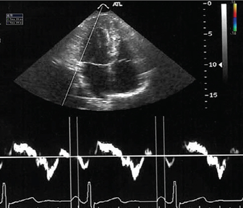
Fig. 1 Measurement of the PA interval with tissue Doppler imaging, which denotes time interval from the onset of P wave on the surface electrocardiogram to the beginning of the late diastolic wave (Am wave). PA: P-wave to the beginning of A-wave on surface ECG.
Cited by 1 articles
-
Evaluation of Left Atrial Electromechanical Delay and Left Atrial Phasic Functions in Surgical Early Menopause Patients
Murat Akcay, Metin Coksevim, Hasan Ulubaşoğlu, Omer Gedikli, Ozcan Yılmaz
J Cardiovasc Imaging. 2019;27(2):137-146. doi: 10.4250/jcvi.2019.27.e22.
Reference
-
1. Arias-Santiago S, Buendía-Eisman A, Aneiros-Fernández J, et al. Lipid levels in patients with lichen planus: a case-control study. J Eur Acad Dermatol Venereol. 2011; 25:1398–1401.2. Arias-Santiago S, Buendía-Eisman A, Aneiros-Fernández J, et al. Cardiovascular risk factors in patients with lichen planus. Am J Med. 2011; 124:543–548.3. Bacaksiz A, Erdogan E, Tasal A, et al. Electrocardiographic P-wave characteristics in patients with psoriasis vulgaris. Ups J Med Sci. 2013; 118:35–41.4. Ahlehoff O, Gislason GH, Jørgensen CH, et al. Psoriasis and risk of atrial fibrillation and ischaemic stroke: a Danish Nationwide Cohort Study. Eur Heart J. 2012; 33:2054–2064.5. Acar G, Sayarlioğlu M, Akçay A, et al. Evaluation of atrial electromechanical delay and left atrial mechanical functions in patients with rheumatoid arthritis. Turk Kardiyol Dern Ars. 2009; 37:447–453.6. Deniz A, Yavuz B, Aytemir K, et al. Intra-left atrial mechanical delay detected by tissue Doppler echocardiography can be a useful marker for paroxysmal atrial fibrillation. Echocardiography. 2009; 26:779–784.7. Deniz A, Sahin DY, Kanadasi M, et al. Conduction characteristics in atrial fibrillation. Predictive value of tissue Doppler echocardiography. Herz. 2014; 39:137–141.8. Sahin M, Bilgili SG, Simsek H, et al. Increased P-wave dispersion in patients with newly diagnosed lichen planus. Clinics (Sao Paulo). 2013; 68:846–850.9. Ari H, Ari S, Akkaya M, et al. Predictive value of atrial electromechanical delay for atrial fibrillation recurrence. Cardiol J. 2013; 20:639–647.10. Simsek H, Gunes Y, Demir C, Sahin M, Gumrukcuoglu HA, Tuncer M. The effects of iron deficiency anemia on p wave duration and dispersion. Clinics (Sao Paulo). 2010; 65:1067–1071.11. Lang RM, Bierig M, Devereux RB, et al. Recommendations for chamber quantification: a report from the American Society of Echocardiography's Guidelines and Standards Committee and the Chamber Quantification Writing Group, developed in conjunction with the European Association of Echocardiography, a branch of the European Society of Cardiology. J Am Soc Echocardiogr. 2005; 18:1440–1463.12. Rudski LG, Lai WW, Afilalo J, et al. Guidelines for the echocardiographic assessment of the right heart in adults: a report from the American Society of Echocardiography endorsed by the European Association of Echocardiography, a registered branch of the European Society of Cardiology, and the Canadian Society of Echocardiography. J Am Soc Echocardiogr. 2010; 23:685–713.13. Sahin M, Simsek H, Akyol A, et al. A new echocardiographic parameter of arterial stiffness in end-stage renal disease. Herz. 2014; 39:749–754.14. Ozer N, Yavuz B, Can I, et al. Doppler tissue evaluation of intra-atrial and interatrial electromechanical delay and comparison with P-wave dispersion in patients with mitral stenosis. J Am Soc Echocardiogr. 2005; 18:945–948.15. Middel P, Lippert U, Hummel KM, et al. Expression of lymphotoxinalpha by keratinocytes: a further mediator for the lichenoid reaction. Pathobiology. 2000; 68:291–300.16. Flammer AJ, Ruschitzka F. Psoriasis and atherosclerosis: two plaques, one syndrome? Eur Heart J. 2012; 33:1989–1991.17. Michelucci A, Bagliani G, Colella A, et al. P wave assessment: state of the art update. Card Electrophysiol Rev. 2002; 6:215–220.18. Calık AN, Ozcan KS, Cağdaş M, et al. Electromechanical delay detected by tissue Doppler echocardiography is associated with the frequency of attacks in patients with lone atrial fibrillation. Cardiol J. 2014; 21:138–143.19. Nar G, İnci S, Aksan G, Soylu K, Demirelli S, Nar R. The relationships between atrial electromechanical delay and CHA2DS2-VASc score in patients diagnosed with paroxysmal AF. Echocardiography. 2015; 32:1359–1366.
