Pulmonary Actinomycosis Imitating Lung Cancer on ¹â¸F-FDG PET/CT: A Case Report and Literature Review
- Affiliations
-
- 1Department of Nuclear Medicine, The First Affiliated Hospital, Sichuan Medical University, Luzhou, Sichuan 64600, China. chenyue5523@126.com
- 2Department of Radiology, The First Affiliated Hospital, Sichuan Medical University, Luzhou, Sichuan 64600, China.
- KMID: 2344280
- DOI: http://doi.org/10.3348/kjr.2015.16.6.1262
Abstract
- Here we report a case of 41-year-old man with a soft tissue density mass at right upper lung and palpable abscesses at right upper backside and right wrist. ¹â¸F-fluorodeoxyglucose positron emission tomography/computed tomography demonstrated a 7.8 × 5.0 cm mass with soft-tissue density in the upper lobe of the right lung with high metabolic activity. The infiltrative mass extended to adjacent chest wall soft tissue. Final diagnosis of pulmonary actinomycosis with multiple abscesses was made. The patient responded well to antibiotics treatment.
Keyword
MeSH Terms
-
Abscess
Actinomycosis/*diagnosis/drug therapy/microbiology
Adult
Anti-Bacterial Agents/therapeutic use
Diagnosis, Differential
Fluorodeoxyglucose F18/chemistry
Humans
Lung Diseases/*diagnosis/drug therapy/microbiology
Lung Neoplasms/pathology
Male
*Positron-Emission Tomography
Tomography, X-Ray Computed
Anti-Bacterial Agents
Fluorodeoxyglucose F18
Figure
-
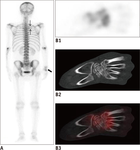
Fig. 1 41-year-old man with pulmonary actinomycosis imitating lung cancer on 99mTc-MDP bone scintigraphy. A. Posterior. B. Uptake of radiotracer is increased at right third and fourth posterior ribs (small black arrows, A) and right multiple carpal bones (large black arrow, A).

Fig. 1 41-year-old man with pulmonary actinomycosis imitating lung cancer with whole body 18F-FDG PET/CT scan. C. Maximum intensity projection showing mass at upper lobe of right lung and peripheral daughter lesions. D. 7.8 × 5.0 cm soft-tissue density mass is demonstrated. Mass has intense 18F-FDG accumulation (SUVmax = 13.3). E, F. Some opaque mottled shadows with 18F-FDG uptake are distributed in multiple lobes of right lung and inferior lobe of left lung. G-I. Infiltrative mass extended to adjacent chest wall, thoracodorsal muscle, and subcutaneous dermal tissues. SUVmax = maximal standardized uptake value, 18F-FDG PET/CT = 18F-fluorodeoxyglucose positron emission tomography/computed tomography
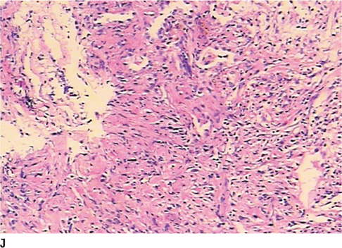
Fig. 1 41-year-old man with pulmonary actinomycosis imitating lung cancer with pathological analysis. J. Pathological result (hematoxylin-eosin staining, × 100) revealed chronic inflammation with proliferation of fibrotic granulation tissue.
Reference
-
1. Arora AK, Nord J, Olofinlade O, Javors B. Esophageal actinomycosis: a case report and review of the literature. Dysphagia. 2003; 18:27–31.2. Mok GS, Choi FP, Chu WC. Actinomycosis imitating parotid cancer with metastatic lymph nodes in FDG PET/CT. Clin Nucl Med. 2011; 36:309–310.3. Ho L, Seto J, Jadvar H. Actinomycosis mimicking anastomotic recurrent esophageal cancer on PET-CT. Clin Nucl Med. 2006; 31:646–647.4. Singla S, Singh H, Mukherjee A, Karunanithi S, Bal C, Kumar R. Cervical and thoracic actinomycosis on (18)F-FDG PET/CT. Clin Nucl Med. 2014; 39:623–624.5. Lim KT, Moon SJ, Kwon JS, Son YW, Choi HY, Choi YY, et al. Urachal actinomycosis mimicking a urachal tumor. Korean J Urol. 2010; 51:438–440.6. Hsu CH, Lee CM, Chia CF, Lin YH. F-18 fluorodeoxyglucose positron emission tomography in an anorectal fistula with actinomycosis. Clin Nucl Med. 2004; 29:452–453.7. Russo TA. Agents of actinomycosis. In : Mandell GL, editor. Principles and practice of infectious disease. 5th ed. Philadelphia: Elsevier, Churchill Livingstone;1995. p. 2645–2654.8. Hoekstra CJ, Hoekstra OS, Teengs JP, Postmus PE, Smit EF. Thoracic actinomycosis imaging with fluorine-18 fluorodeoxyglucose positron emission tomography. Clin Nucl Med. 1999; 24:529–530.9. Kanda H, Nakamura Y, Nagata T, Fukumori K, Imoto Y, Tabata K, et al. [Surgery for pulmonary actinomycosis that was difficult to differentiate from lung cancer; report of a case]. Kyobu Geka. 2011; 64:864–867.10. Kogure S, Yamamoto N, Watanabe F, Yuasa U, Tokui T, Shomura S. [Pulmonary actinomycosis which was clinically suggested lung cancer; report of a case]. Kyobu Geka. 2011; 64:254–257.11. Okuda R, Izumo T, Yoshikawa M, Kakuta Y, Tamaoki J, Nagai A. [A case of thoracic actinomycosis in the left lung coexisting with pulmonary squamous cell carcinoma in the right lung]. Nihon Kokyuki Gakkai Zasshi. 2011; 49:103–107.12. Taştepe AI, Ulaşan NG, Liman ST, Demircan S, Uzar A. Thoracic actinomycosis. Eur J Cardiothorac Surg. 1998; 14:578–583.13. Yeung VH, Wong QH, Chao NS, Leung MW, Kwok WK. Thoracic actinomycosis in an adolescent mimicking chest wall tumor or pulmonary tuberculosis. Pediatr Surg Int. 2008; 24:751–754.
- Full Text Links
-
- Actions
-
Cited
- CITED
-
- Close
- Share
-
- Similar articles
-
- Role of ¹â¸F-FDG PET-CT in Monitoring the Cyclophosphamide Induced Pulmonary Toxicity in Patients with Breast Cancer: 2 Case Reports
- Comparison of Neck CT and ¹â¸F-FDG PET-CT for Making the Preoperative Diagnosis of Lymph Node Metastasis in Papillary Thyroid Cancer
- â¶â¸Gallium-Arginine-Glycine-Aspartic Acid and ¹â¸F-Fluorodeoxyglucose Positron Emission Tomography/Computed Tomography in Chondroblastic Osteosarcoma of the Skull
- ¹â¸F-FDG PET/MR Refines Evaluation in Newly Diagnosed Metastatic Urethral Adenocarcinoma
- [18F]FDG PET/CT Findings of Mass‑Forming Actinomycosis in an Uncontrolled Diabetic Patient
