Extent of Disc Degeneration after Single-Level Cervical Anterior Microforaminotomy Analyzed with Long-Term Radiological Data
- Affiliations
-
- 1Department of Emergency Medicine, School of Medicine, Ewha Womans University, Seoul, Korea.
- 2Department of Neurosurgery, School of Medicine, Ewha Womans University, Seoul, Korea. kimmh@ewha.ac.kr
- KMID: 2339957
- DOI: http://doi.org/10.3340/jkns.2014.56.3.200
Abstract
OBJECTIVE
To prove the extents and details of cervical degeneration after anterior microforaminotomy (AMF) with 6-years follow-up.
METHODS
A retrospective study of 24 patients, underwent single-level AMF, was performed. Clinical and radiologic data were analyzed with office charts, questionaires, and picture achieving and communication system images.
RESULTS
According to Odom's criteria, 91.6% achieved favorable outcome. The mean visual analog scale score was improved from 8.6 to 3, and the mean neck disability index was improved from 27.9 to 7.3 (p<0.01). Eighteen cases (75%) showed disc height (DH) decrease. The disc invasion was correlated with DH decrease (p<0.05). The disc height decrease correlated with static, dynamic changes of shell angle and spur formation (p<0.05). Any radiological parameters did not affect the clinical outcome.
CONCLUSION
AMF is an effective technique for treating unilateral cervical radiculopathy. It showed excellent surgical outcomes even in long-term follow-ups. However, a decrease in DH occurred in a considerable number of patients. Disc invasion during surgery may be the trigger of sequential degeneration.
MeSH Terms
Figure
-
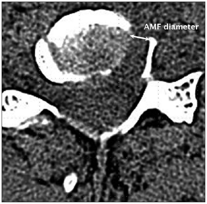
Fig. 1 Measurement of diameter of anterior microforaminotomy on axial CT image. AMF : anterior microforaminotomy.
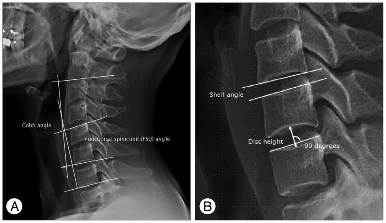
Fig. 2 Measurement of static angles on plain cervical lateral image. A : Functional spine unit and Cobb angles. B : Measurement of disc height and shell angle on a plain radiograph in neutral lateral position.
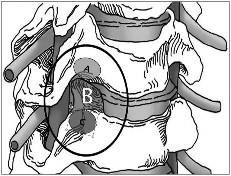
Fig. 3 Illustration showing the various entry and resection sites of microforaminotomy. A : Entry point of upper transcorporeal approach. B : Resection site of transuncal approach. C : Entry point of lower transcorporeal approach.
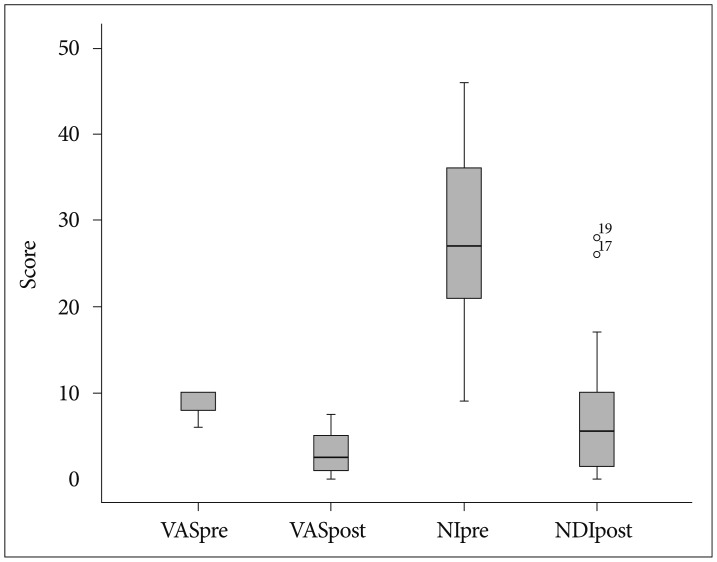
Fig. 4 Box-plot graph showing preoperative and postoperative changes of visual analogue scale (VAS) and neck disability index (NDI) scores with mean, maximum, and minimum values. VASpre : preoperative VAS, VASpost : postoperative VAS, NDIpre : preoperative NDI, NDIpost : postoperative NDI.
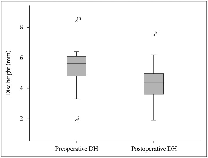
Fig. 5 Box-plot graph showing changes in preoperative and postoperative disc height (DH) with mean, maximum, and minimum values.
Reference
-
1. Balasubramanian C, Price R, Brydon H. Anterior cervical microforaminotomy for cervical radiculopathy--results and review. Minim Invasive Neurosurg. 2008; 51:258–262. PMID: 18855288.
Article2. Choi G, Lee SH, Bhanot A, Chae YS, Jung B, Lee S. Modified transcorporeal anterior cervical microforaminotomy for cervical radiculopathy : a technical note and early results. Eur Spine J. 2007; 16:1387–1393. PMID: 17203272.
Article3. Cornelius JF, Bruneau M, George B. Microsurgical cervical nerve root decompression via an anterolateral approach : clinical outcome of patients treated for spondylotic radiculopathy. Neurosurgery. 2007; 61:972–980. discussion 980. PMID: 18091274.
Article4. Cuellar VG, Cuellar JM, Vaccaro AR, Carragee EJ, Scuderi GJ. Accelerated degeneration after failed cervical and lumbar nucleoplasty. J Spinal Disord Tech. 2010; 23:521–524. PMID: 21131800.
Article5. Hong WJ, Kim WK, Park CW, Lee SG, Yoo CJ, Kim YB, et al. Comparison between transuncal approach and upper vertebral transcorporeal approach for unilateral cervical radiculopathy - a preliminary report. Minim Invasive Neurosurg. 2006; 49:296–301. PMID: 17163344.
Article6. Hussain M, Natarajan RN, An HS, Andersson GB. Patterns of height changes in anterior and posterior cervical disc regions affects the contact loading at posterior facets during moderate and severe disc degeneration : a poroelastic C5-C6 finite element model study. Spine (Phila Pa 1976). 2010; 35:E873–E881. PMID: 21289493.7. Jho HD. Microsurgical anterior cervical foraminotomy for radiculopathy : a new approach to cervical disc herniation. J Neurosurg. 1996; 84:155–160. PMID: 8592215.
Article8. Jho HD, Kim WK, Kim MH. Anterior microforaminotomy for treatment of cervical radiculopathy : part 1--disc-preserving "functional cervical disc surgery". Neurosurgery. 2002; 51(5 Suppl):S46–S53. PMID: 12234429.9. Johnson JP, Filler AG, McBride DQ, Batzdorf U. Anterior cervical foraminotomy for unilateral radicular disease. Spine (Phila Pa 1976). 2000; 25:905–909. PMID: 10767800.
Article10. Kirkaldy-Willis WH, Farfan HF. Instability of the lumbar spine. Clin Orthop Relat Res. 1982; (165):110–123. PMID: 6210480.
Article11. Kotil K, Bilge T. Prospective study of anterior cervical microforaminotomy for cervical radiculopathy. J Clin Neurosci. 2008; 15:749–756. PMID: 18378143.
Article12. Kumaresan S, Yoganandan N, Pintar FA, Maiman DJ, Goel VK. Contribution of disc degeneration to osteophyte formation in the cervical spine : a biomechanical investigation. J Orthop Res. 2001; 19:977–984. PMID: 11562150.
Article13. Lee JY, Löhr M, Impekoven P, Koebke J, Ernestus RI, Ebel H, et al. Small keyhole transuncal foraminotomy for unilateral cervical radiculopathy. Acta Neurochir (Wien). 2006; 148:951–958. PMID: 16804642.
Article14. Miyazaki M, Hymanson HJ, Morishita Y, He W, Zhang H, Wu G, et al. Kinematic analysis of the relationship between sagittal alignment and disc degeneration in the cervical spine. Spine (Phila Pa 1976). 2008; 33:E870–E876. PMID: 18978580.
Article15. Nassr A, Lee JY, Bashir RS, Rihn JA, Eck JC, Kang JD, et al. Does incorrect level needle localization during anterior cervical discectomy and fusion lead to accelerated disc degeneration? Spine. 2009; 34:189–192. PMID: 19139670.
Article16. Osti OL, Vernon-Roberts B, Fraser RD. 1990 Volvo Award in experimental studies. Anulus tears and intervertebral disc degeneration. An experimental study using an animal model. Spine (Phila Pa 1976). 1990; 15:762–767. PMID: 2237626.
Article17. Saringer W, Nöbauer I, Reddy M, Tschabitscher M, Horaczek A. Microsurgical anterior cervical foraminotomy (uncoforaminotomy) for unilateral radiculopathy : clinical results of a new technique. Acta Neurochir (Wien). 2002; 144:685–694. PMID: 12181702.18. Schendel MJ, Wood KB, Buttermann GR, Lewis JL, Ogilvie JW. Experimental measurement of ligament force, facet force, and segment motion in the human lumbar spine. J Biomech. 1993; 26:427–438. PMID: 8478347.
Article19. Tanaka N, An HS, Lim TH, Fujiwara A, Jeon CH, Haughton VM. The relationship between disc degeneration and flexibility of the lumbar spine. Spine J. 2001; 1:47–56. PMID: 14588368.
Article
- Full Text Links
-
- Actions
-
Cited
- CITED
-
- Close
- Share
-
- Similar articles
-
- Does the Size of Anterior Microforaminotomy Affect the Surgical Outcome?
- The Outcome of Anterior Microforaminotomy for Single Level Cervical Radicular Disease
- The Result of Posterior Microforaminotomy for Posterolateral Herniation of Cervical Discs
- Anterior Cervical Microforaminotomy
- Midterm Follow-up Results of Anterior Cervical Microforaminotomy
