A Case of Orbital Organizing Hematoma Presenting as a Chalazion
- Affiliations
-
- 1Department of Ophthalmology, Hallym University College of Medicine, Anyang, Korea. tweeti2@hanmail.net
Abstract
- PURPOSE
Organizing hematomas (hematic pseudocysts) of the orbit are usually the consequence of direct blunt trauma and are important in the differential diagnosis of orbital cystic lesion. Herein, we report a case of orbital organizing hematoma masquerading as a chalazion.
CASE SUMMARY
A 12-year-old female visited our clinic complaining of left lower eyelid swelling. CT scan and MR imaging showed a mass detected in the inferomedial space of her left orbit, which did not invade the adjacent tissue. Excisional biopsy of the orbital mass was performed. Histological examination showed the accumulation of blood-breakdown products within a thick fibrous capsule without epithelial or endothelial lining. The mass was diagnosed as an organizing hematoma.
CONCLUSIONS
An orbital organizing hematoma can present, although rare, as a chalazion-like eyelid mass. Careful clinical and radiological examinations can help in making a differential diagnosis.
Keyword
MeSH Terms
Figure
-

Figure 1. (A, B) External photographs show left lower eyelid swelling and partially protruded mass. At 1 month after surgery, the patient has no evidence of recurrence.
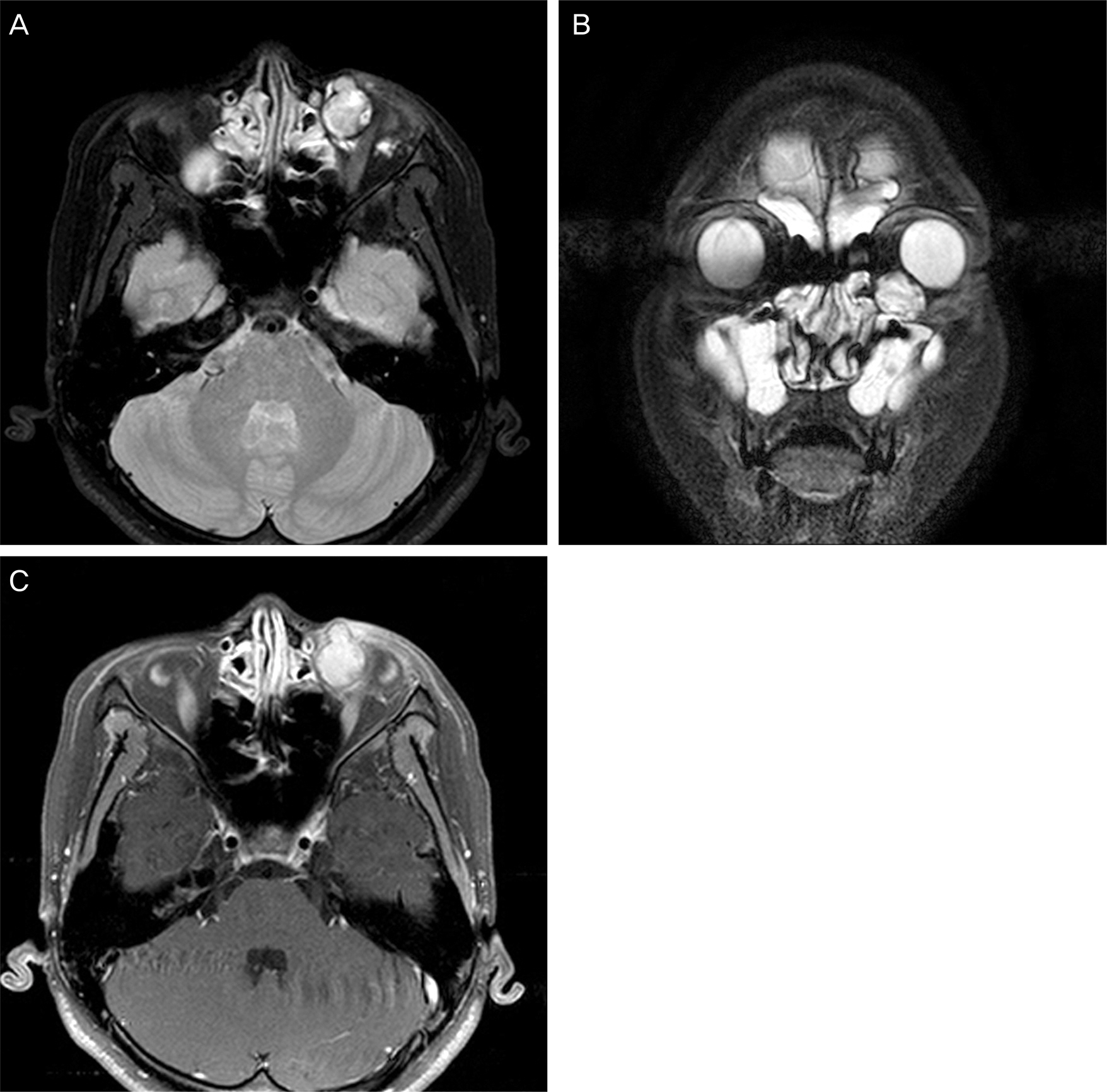
Figure 2. Axial view of T1-weighted image (A) and co-ronal view of T2-weighted image (B) demonstrate 1.7 ×1.7 cm sized well-demarcated cystic mass in the inferior medial extraconal space of the left orbit. The mass shows high signal intensity in both T1 and T2-weighted images. There is maxillary sinusitis. (C) The mass shows poor enhancement.
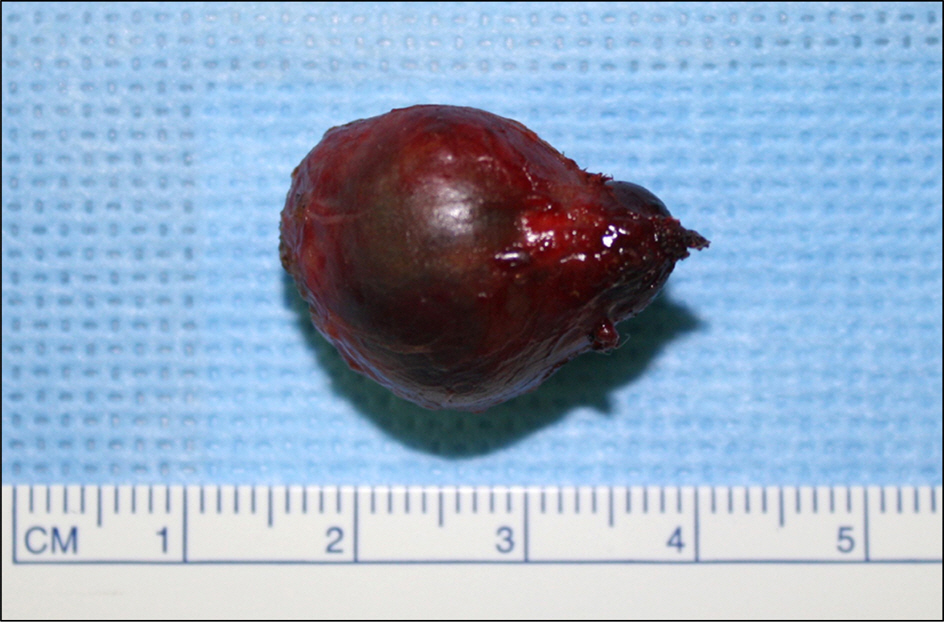
Figure 3. The gross appearance of the excised mass. The mass showed dark reddish color and had smooth surface. The size was 25 × 18 × 15 mm.
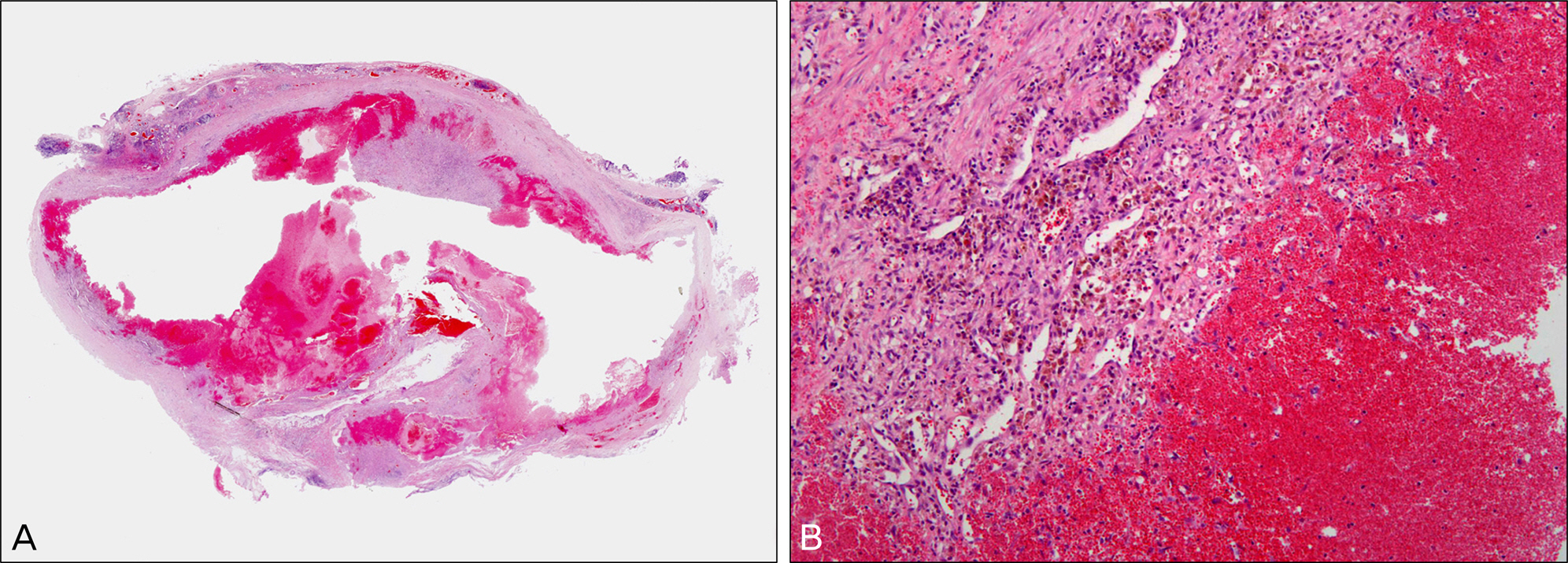
Figure 4. Microscopic appearance of organizing hematoma (A) The tumor was composed of thick fibrous capsule lacking an epithelial lining, and blood clots (H&E, ×12.5). (B) The blood-breakdown products and inflammatory cells are observed. (H&E, ×100).
Reference
-
References
1. Atalla ML, McNab AA, Sullivan TJ, Sloan B. Nontraumatic sub-periosteal orbital hemorrhage. Ophthalmology. 2001; 108:183–9.
Article2. Huh KC, Kim YD. A case of subperiosteal orbital hematoma asso-ciated with von willebrand disease. J Korean Opthalmol Soc. 2002; 43:213–8.3. Nerea SM, Lgnacio AG, Pilar TP, Ana RG. Spontaneous sub-periosteal hematoma of the orbit. J Pediatr Ophthalmol Strabismus. 2009; 46:175–7.
Article4. Woo KI, Kim YD. Subperiosteal hematoma of the orbit associated with sinusitis. Korean J Ophthalmol. 1997; 11:118–22.
Article5. Guirgis MF, Segal WA, Lueder GT. Subperiosteal orbital hemor-rhage as initial manifestation of Christmas disease (factor IX defi-ciency). Am J Ophthalmol. 2002; 133:584–5.
Article6. Goldberg SH, Sassani JW, Parnes RE. Traumatic intraconal hem-atic cyst of the orbit. Arch Ophthalmol. 1992; 110:378–80.
Article7. Yoshikawa K, Fujisawa H, Kajiwara K. . Cause of hematic cysts of the orbit: increased fibrinolysis and immunohistologic expression of tissue plasminogen activator. Ophthalmology. 2009; 116:130–4.
Article8. Dobben GD, Philip B, Mafee MF. . Orbital subperiosteal hem-atoma, cholesterol granuloma, and infection. Evaluation with MR imaging and CT. Radiol Clin North Am. 1998; 36:1185–200.
Article9. Park HW, Lee BJ, Chung YS. Orbital subperiosteal hematoma as-sociated with sinus infection. Rhinology. 2010; 48:117–22.
Article10. Lee JJ, Kim JH, Hwang SW. . A case of intraconal hematic pseudocyst. J Korean Ophthalmol Soc. 2006; 47:661–6.11. Katowitz WR, Shields CL, Shields JA. . Pilomatrixoma of the eyelid simulating a chalazion. J Pediatr Ophthalmol Strabismus. 2003; 40:247–8.
Article12. Maheshwari R, Maheshwari S. Extramedullary plasmacytoma masquerading as chalazion. Orbit. 2009; 28(2-3):191–3.13. Hui JI, Buchser NM, Dubovy SR. Primary eyelid leiomyoma. Ophthal Plast Reconstr Surg. 2011; 27:e102–3.
Article14. Rahimi M, Moinfar N, Ashrafi A. Eyelid leishmaniasis masquer-ading as chalazia. Eye (Lond). 2009; 23:737.
Article
- Full Text Links
-
- Actions
-
Cited
- CITED
-
- Close
- Share
-
- Similar articles
-
- A Case of Organizing Hematoma of the Nasal Septum
- Three Cases of Organizing Hematoma in the Maxillary Sinus
- A Case of Surgical Management for Orbital Organizing Hematoma from Orbital Varix
- Non-Traumatic Organizing Hematoma on Supraclavicular Fossa in a Child: A Case Report
- A Case of Orbital Hematoma Caused by Traumatic Parietal Subperiosteal Hemorrhage
