Considering Spherical Aberration in Choosing the Wavefront Map for Laser Vision Correction
- Affiliations
-
- 1Department of Ophthalmology, Dongguk University, College of Medicine, Gyeongju, Korea.
- 2Department of Ophthalmology, Dongguk University, Graduate School of Medicine, Goyang, Korea. cpark@duih.org
- 3Department of Ophthalmology, Dongguk University, Ilsan Hospita, Goyang, Korea.
Abstract
- PURPOSE
To report the dynamic nature of human optical aberrations in the scotopic condition.
METHODS
A total of 20 eyes who were candidates for laser vision correction were included in the present study. Repeated wavefront data were obtained using WavescanTM (AMO/VISX). From the wavefront analysis data, the sphere, astigmatism, average pupil size, spherical aberration, coma and trefoil were selected and used to investigate any correlation among the parameters.
RESULTS
The sphere, spherical aberration, coma and pupil size showed a dynamic change in the scotopic condition. The spherical aberration and pupil size decreased by the amount of 0.10 +/- 0.04 microm and 0.55 +/- 0.37 mm as the sphere changed 1 D in myopic direction. There was significant positive correlation between the sphere and spherical aberration in 13 eyes of 9 patients (65%), between the sphere and pupil size in 5 eyes of 4 patients (25%), and between the sphere and coma in 3 eyes of 3 patients (15%). The spherical aberration decreased significantly in 4 eyes of 4 patients (20%) as the pupil size decreased.
CONCLUSIONS
The optical aberration of human eyes showed a dynamic nature in the scotopic condition. In particular, there was significant correlation between the sphere and spherical aberration. The observed correlations have the potential to be used as helpful indicators to select the optimal wavefront data for the laser vision correction.
Figure
-

Figure 1. The correlation between the change in sphere (Δ sphere) and the change in spherical aberration (Δ SA), (A), and the correlation between the change in sphere (Δ sphere) and the change of pupil size (Δ pupil), (B), were demonstrated. The spherical aberration decreased on average of 0.10 ± 0.04 μ m (mean ± SD) as the sphere decreased 1 diopter (rho=0.747, p<0.001, Spearman's correlation test). The pupil size decreased on average 0.55 ± 0.37 mm as the sphere decreased 1 diopter (rho=0.526, p=0.017, Spearman's correlation test).
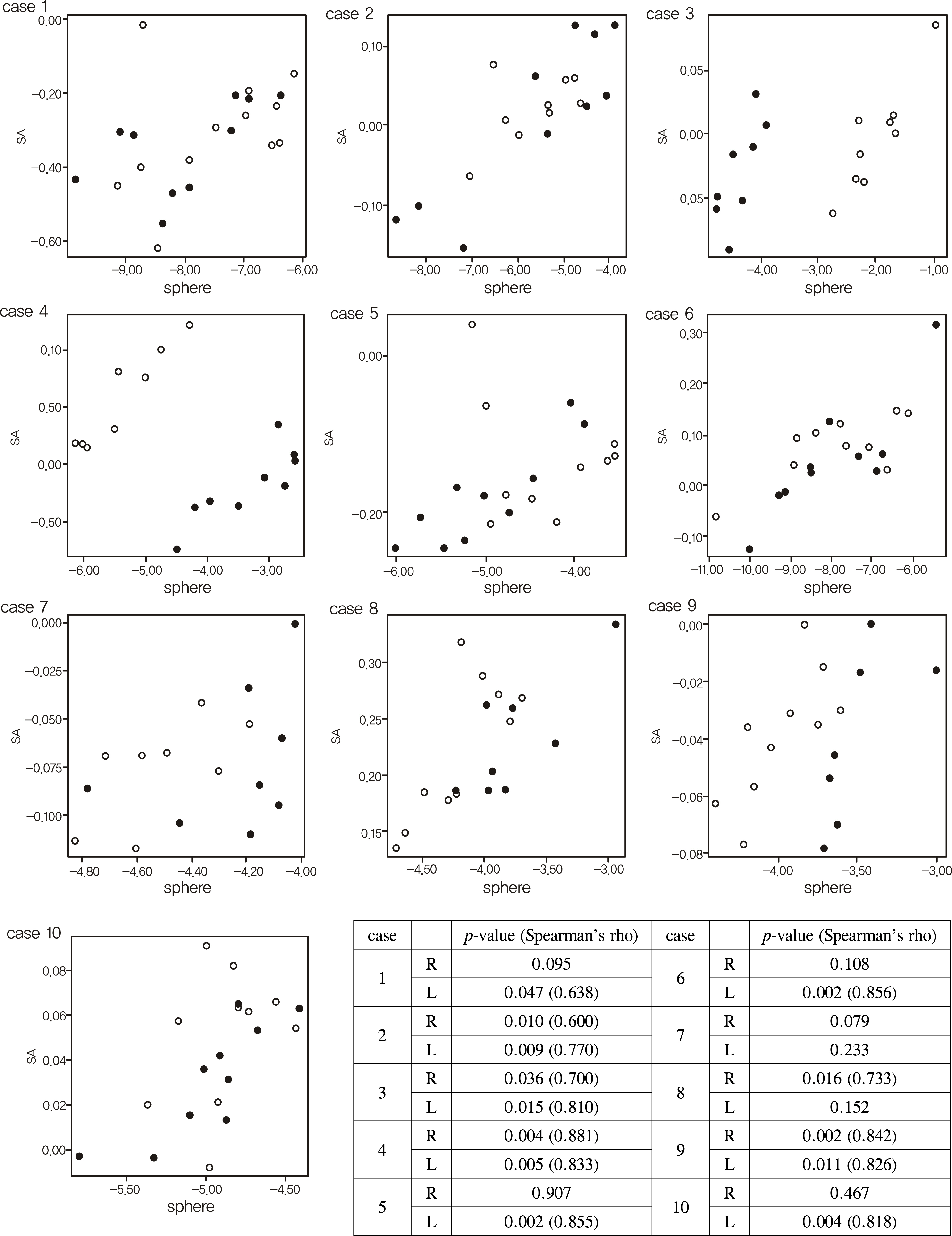
Figure 2. The change in spherical aberration according to the change in sphere in study eyes. The scatter plot showed that 13 eyes of 9 patients showed significant correlation between SA and sphere (refer to inserted table for Spearman's rho and p-value). Filled dots mean the values of the left eye and empty dots mean the values of the right eye.
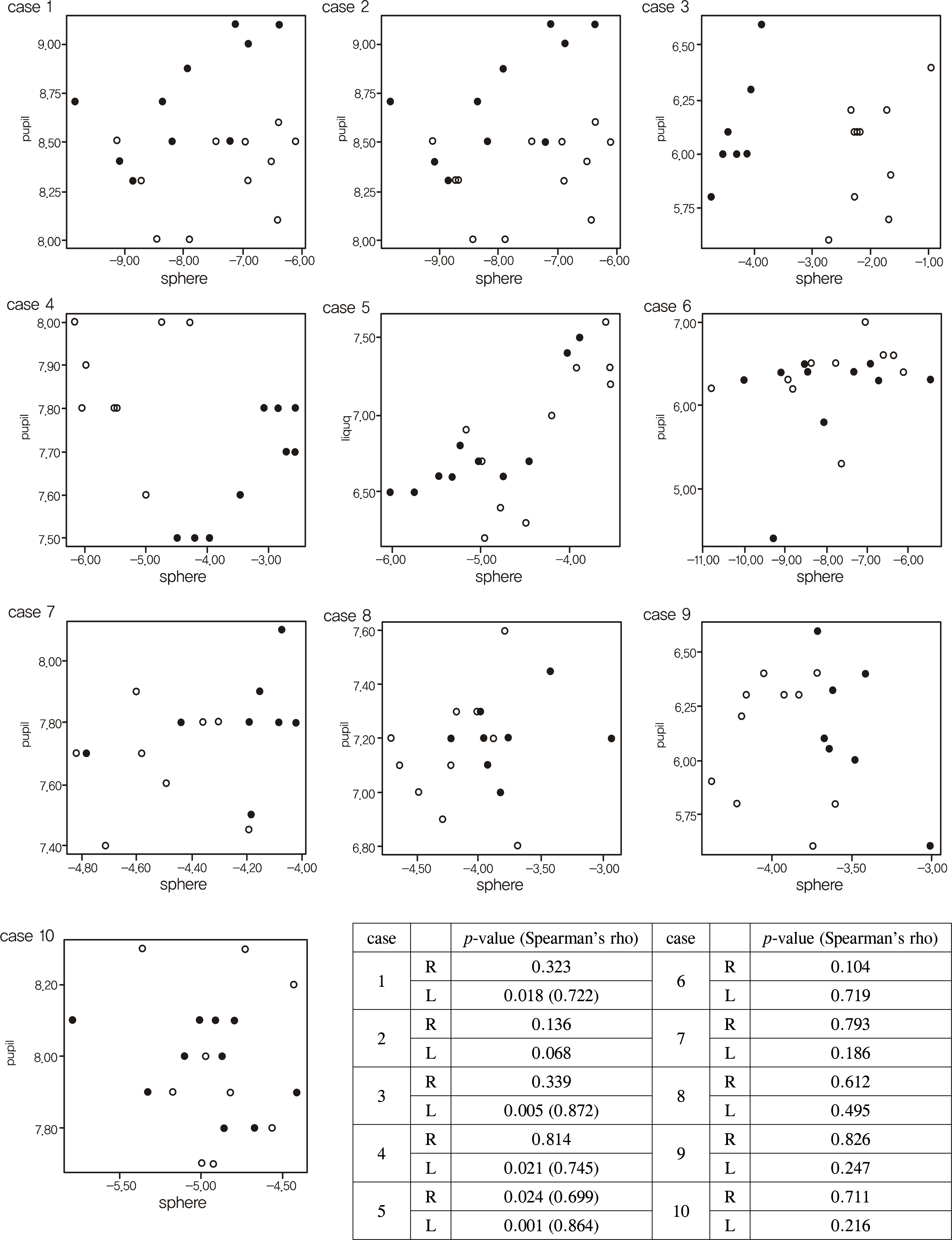
Figure 3. The change in pupil size according to the change in sphere in study eyes. The scatter plot showed that 5 eyes of 4 patients showed significant correlation between pupil size and sphere (refer to inserted table for Spearman's rho and p-value). Filled dots mean the values of the left eye and empty dots mean the values of the right eye.
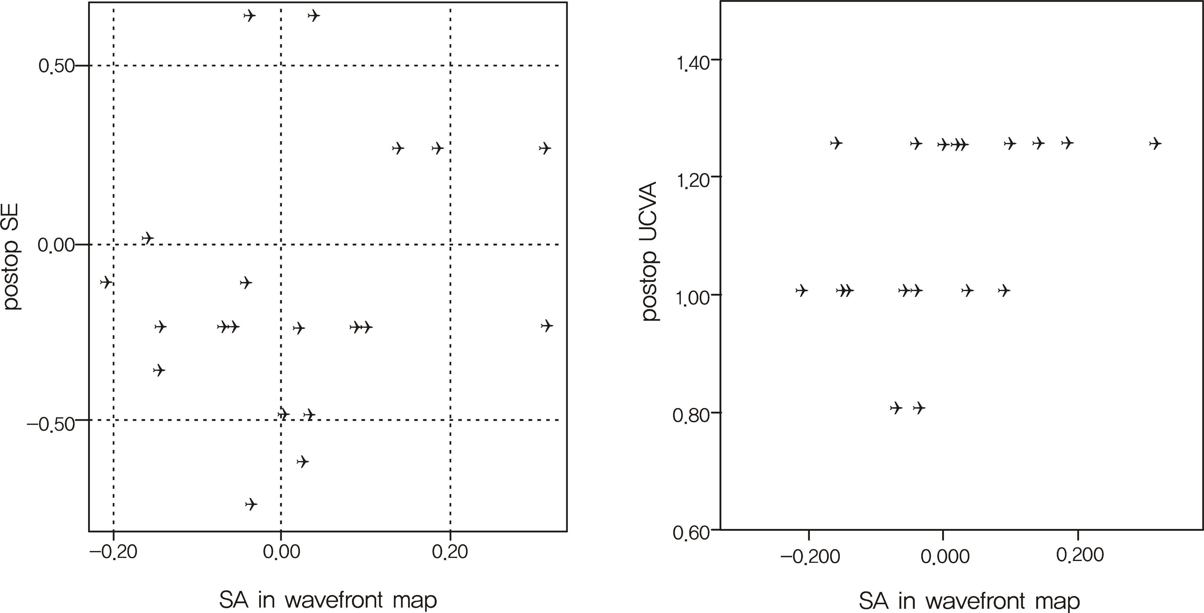
Figure 4. The correlation between the SA used for the laser vision correction and the postoperative spherical equivalent was not significant (p=0.510, Spearman's correlation test). However, UCVA showed the positive correlation with SA (p=0.039, rho=0.464, Spearman's correlation test).
Reference
-
References
1. Charman WN. Wavefront aberration of the eye: a review. Optom Vis Sci. 1991; 68:574–83.2. Maeda N. Wavefront technology in ophthalmology. Curr Opin Ophthalmol. 2001; 12:294–9.
Article3. Oshika T, Klyce SD, Applegate RA, et al. Comparison of corneal wavefront aberrations after photorefractive keratectomy and laser in situ keratomileusis. Am J Ophthalmol. 1999; 127:1–7.
Article4. Awwad ST, Bowman RW, Cavanagh HD, McCulley JP. Wavefront-guided LASIK for myopia using the LADAR CustomCornea and the VISX CustomVue. J Refract Surg. 2007; 23:26–38.
Article5. Subbaram MV, MacRae SM. Does dilated wavefront aberration measurement provide better postoperative outcome after custom LASIK? Ophthalmology. 2006; 113:1813–7.
Article6. Netto MV, Dupps W Jr, Wilson SE. Wavefront-guided ablation: evidence for efficacy compared to traditional ablation. Am J Ophthalmol. 2006; 141:360–8.
Article7. Thibos LN, Applegate RA, Schwiegerling JT, Webb R; VSIA Standards Taskforce Members. Standards for reporting the optical aberrations of eyes. J Refract Surg. 2002; 18:S652–60.8. Yoon G, Jeong TM, Cox IG, Williams DR. Vision improvement by correcting higher-order aberrations with phase plates in normal eyes. J Refract Surg. 2004; 20:S523–7.
Article9. Applegate RA, Sarver EJ, Khemsara V. Are all aberrations equal? J Refract Surg. 2002; 18:S556–62.
Article10. Guirao A, Williams DR. A method to predict refractive errors from wave aberration data. Optom Vis Sci. 2003; 80:36–42.
Article11. Netto MV, Ambrósio R Jr, Shen TT, Wilson SE. Wavefront analysis in normal refractive surgery candidates. J Refract Surg. 2005; 21:332–8.
Article12. Yoon G, Macrae S, Williams DR, Cox IG. Causes of spherical aberration induced by laser refractive surgery. J Cataract Refract Surg. 2005; 31:127–35.
Article13. Roberts C. Biomechanics of the cornea and wavefront-guided laser refractive surgery. J Refract Surg. 2002; 18:S589–92.
Article14. Kulkamthorn T, Silao JN, Torres LF, et al. Wavefront-guided laser in situ keratomileusis in the treatment of high myopia by using the CustomVue wavefront platform. Cornea. 2008; 27:787–90.
Article15. Perez-Straziota CE, Randleman JB, Stulting RD. Visual acuity and higher-order aberrations with wavefront-guided and wavefront- optimized laser in situ keratomileusis. J Cataract Refract Surg. 2010; 36:437–41.16. Reinstein DZ, Neal DR, Vogelsang H, et al. Optimized and wave-front guided corneal refractive surgery using the Carl Zeiss Meditec platform: the WASCA aberrometer, CRS-Master, and MEL80 excimer laser. Ophthalmol Clin North Am. 2004; 17:191–210.
Article17. Venter J. Wavefront-guided custom ablation for myopia using the NIDEK NAVEX laser system. J Refract Surg. 2008; 24:487–93.
Article18. Cheng H, Barnett JK, Vilupuru AS, et al. A population study on changes in wave aberrations with accommodation. J Vis. 2004; 4:272–80.
Article19. Iida Y, Shimizu K, Ito M, Suzuki M. Influence of age on ocular wavefront aberration changes with accommodation. J Refract Surg. 2008; 24:696–701.
Article20. He JC, Burns SA, Marcos S. Monochromatic aberrations in the ac-commodated human eye. Vision Res. 2000; 40:41–8.
Article21. Dubbelman M, Van der Heijde GL, Weeber HA. Change in shape of the aging human crystalline lens with accommodation. Vision Res. 2005; 45:117–32.
Article22. Dubbelman M, Van der Heijde GL, Weeber HA, Vrensen GF. Changes in the internal structure of the human crystalline lens with age and accommodation. Vision Res. 2003; 43:2363–75.
Article23. Ninomiya S, Fujikado T, Kuroda T, et al. Changes of ocular aberration with accommodation. Am J Ophthalmol. 2002; 134:924–6.
Article24. Seiler T, Koller T. Asphericity of the cornea and astigmatism. Klin Monbl Augenheilkd. 2005; 222:977–82.25. Rosen ES, Eustace P, Thompson HS, Cumming WJK. NeuroOphthalmology. London: Mosby;1998. p. 33–4.
- Full Text Links
-
- Actions
-
Cited
- CITED
-
- Close
- Share
-
- Similar articles
-
- Change of High-order Aberration after Wavefront-guided LASIK and LASEK
- Higher-Order Aberrations and Visual Acuity with Wavefront-Guided and Wavefront-Optimized Ablation in Laser Keratorefractive Surgery
- Analysis of Higher-Order Wavefront Aberrations in Standard PRK
- Age and Spherical Equivalent Related Changes in Wavefront Aberrations
- Clinical Result of Wavefront-guided Corneal Ablation: LASIK vs. LASEK
