J Korean Ophthalmol Soc.
2010 Mar;51(3):333-339.
Comparison of Visual Function Between Two Aspheric Intraocular Lenses After Microcoaxial Cataract Surgery
- Affiliations
-
- 1Department of Ophthalmology, Chonnam National University Medical School and Hospital, Gwangju, Korea. kcyoon@chonnam.ac.kr
Abstract
- PURPOSE
To compare the visual function and postoperative refraction between two aspheric intraocular lenses (IOLs) after microcoaxial cataract surgery.
METHODS
A prospective analysis of 60 eyes of 58 patients who had undergone microcoaxial phacoemulsification and implantation of aspheric IOLs (AcrySof IQ, 30 eyes; Akreos MI60, 30 eyes) was performed. The best corrected visual acuity (BCVA), spherical equivalent, intraocular pressure (IOP), corneal thickness, endothelial cell density (ECD), predictability of postoperative spherical equivalent, higher order aberrations, contrast sensitivity test, satisfaction, and glare were evaluated during the follow-up period of three months.
RESULTS
There were no significant differences in BCVA, spherical equivalent, IOP, corneal thickness, or ECD (p>0.05). The predictability of the postoperative spherical equivalent in the AcrySof IQ and Akreos MI60 IOL groups was not significantly different (p=0.59), and the two groups showed good anterior-posterior stability during the postoperative three months. There were no significant differences in higher order aberrations, contrast sensitivity test, satisfaction or glare (p>0.05).
CONCLUSIONS
Both AcrySof IQ and Akreos MI60 implantation groups showed similar visual functions and postoperative spherical equivalents after microcoaxial cataract surgery.
Keyword
MeSH Terms
Figure
-
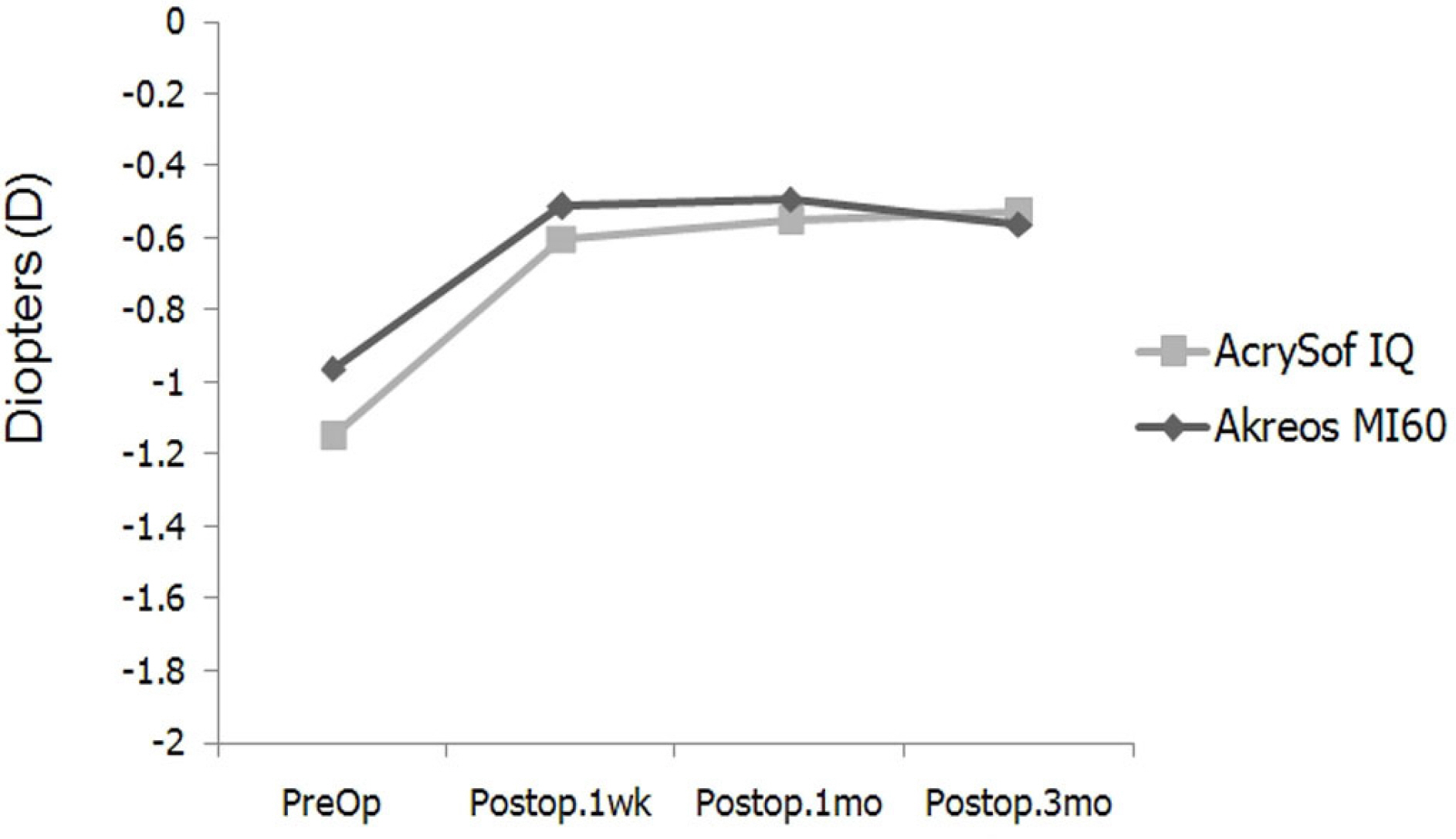
Figure 1. Changes in the spherical equivalent after microcoaxial cataract surgery in the two aspheric intraocular lenses groups.
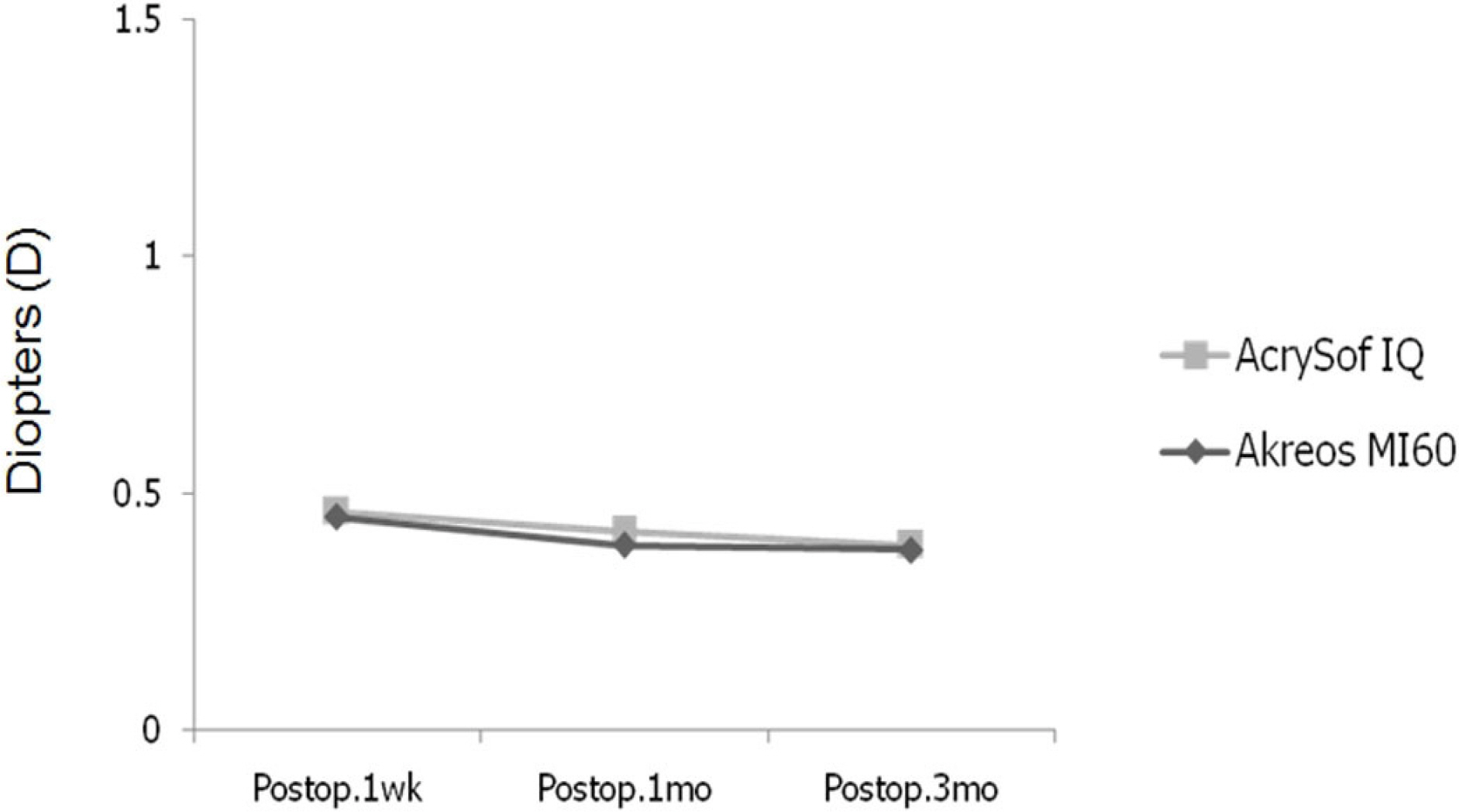
Figure 2. Changes in the surgically induced astigmatism in the two aspheric intraocular lenses groups.
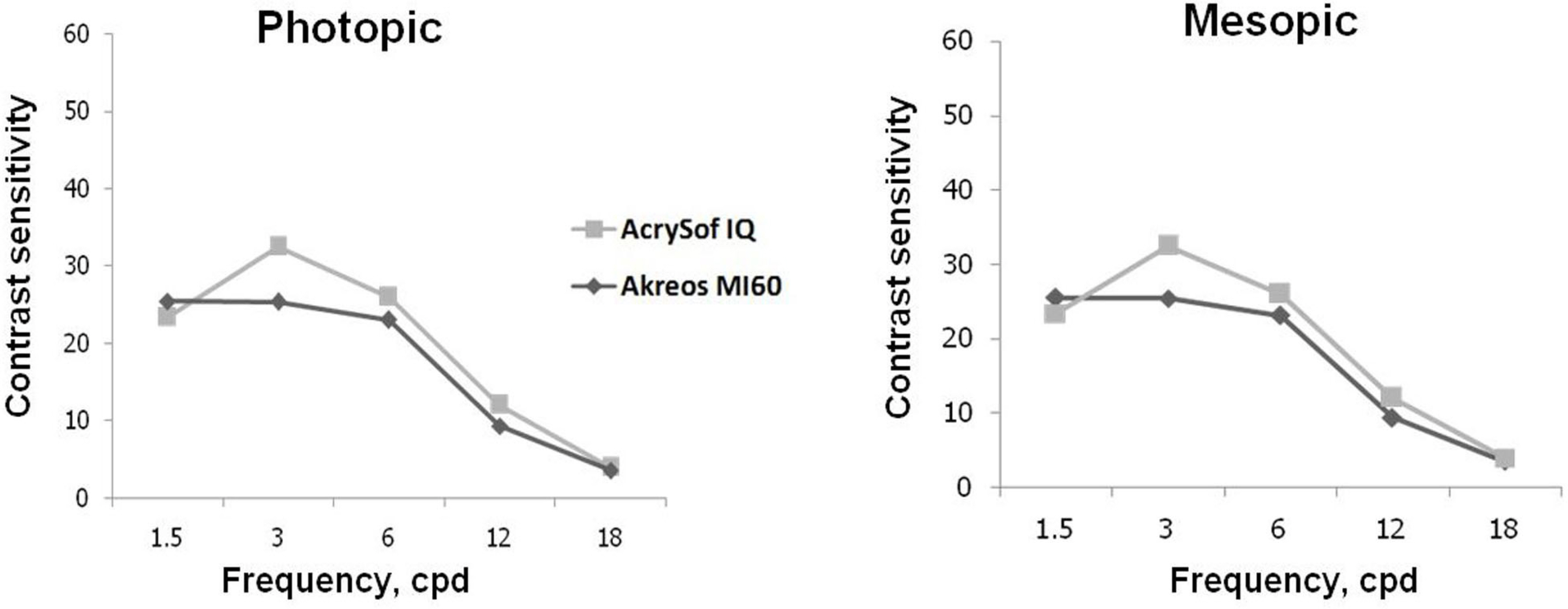
Figure 3. Comparison of contrast sensitivity under photopic condition and mesopic condition three months postoperatively.
Reference
-
References
1. Kellman CD. abdominal and aspiration; a new technique of cataract removal; a preliminary report. Am J Ophthalmol. 1967; 64:23–35.2. Vasavada V, Vasavada V, Raj SM, Vasavada AR. Intraoperative performance and postoperative outcomes of microcoaxial phacoemulsification. Observational study. J Cataract Refract Surg. 2007; 33:1019–24.3. Oscher RH, Injev VP. Microcoaxial phacoemulsification Part 1: laboratory studies. J Cataract Refract Surg. 2007; 33:401–7.4. Oscher RH. Microcoaxial phacoemulsification Part 2: clinical studies. J Cataract Refract Surg. 2007; 33:408–12.5. Werner L, Mamalis N. Wavefront corrections of intraocular lenses. Ophthalmol Clin North Am. 2004; 17:233–45.
Article6. Chalita MR, Krueger RR. Correlation of aberrations with visual acuity and symptoms. Ophthalmol Clin North Am. 2004; 17:135–42.
Article7. Guirao A, Redondo M, Geraghty E, et al. Corneal optical aberrations and retinal image quality in patients whom monofocal intraocular lenses were implanted. Arch Ophthalmol. 2002; 120:1143–51.8. Holladay JT, Moran JR, Kezirian GM. Analysis of aggregate surgically induced refractive change, prediction error, and intraocular astigmatism. J Cataract Refract Surg. 2002; 28:206–7.
Article9. Huang FC, Tseng SH. Comparison of surgically induced astigmatism after sutureless temporal clear corneal and scleral frown incisions. J Cataract Refract Surg. 1998; 24:477–81.
Article10. Lee DS, Joo CK. Effect of incision length on visual recovery and astigmatism in no-suture cataract surgery. J Korean Ophthalmol Soc. 1992; 33:470–5.11. Hu YJ, Joo CK. Surgically induced astigmatism after temporal clear corneal incision in sutureless cataract surgery. J Korean Ophthalmol Soc. 1998; 39:2622–7.12. Jee DH, Lee PY, Joo CK. The comparison of astigmatism according to the incision size in cataract operation. J Korean Ophthalmol Soc. 2003; 44:594–8.13. Kim HJ, Kim JH, Lee DH. Endothelial cell damage in microincision cataract surgery and coaxial phacoemulsification. J Korean Ophthalmol Soc. 2007; 48:1630–5.14. Cavallini GM, Campi L, Masini C, et al. Bimanual microphacoemulsification vs coaxial miniphacoemulsification: Prospective study. J Cataract Refract Surg. 2007; 33:387–92.15. Choi JA, Chung SK, Kim HS. Comparative Study of Microcoaxial Cataract Surgery and Conventional Cataract Surgery. J Korean Ophthalmol Soc. 2008; 49:904–10.
Article16. Hayashi K, Hayashi H, Nakao F, Hayashi F. Risk factors for corneal endothelial injury during phacoemulsification. J Cataract Refract Surg. 1996; 22:1079–84.
Article17. Dick HB, Kohnen T, Jacobi FK, Jacobi KW. abdominal endothelial cell loss following phacoemulsification through a temporal clear corneal incision. J Cataract Refract Surg. 1996; 22:63–71.18. Joussen AM, Barth U, Cubuk H, Koch H. Effect of irrigating abdominal and irrigation temperature on the cornea and pupil during phacoemulsification. J Cataract Refract Surg. 2000; 26:392–7.19. Bourne RR, Minassian DC, Dart JK, et al. Effect of cataract abdominal on the corneal endothelium; modern phacoemulsification compared with extracapsular cataract surgery. Ophthalmology. 2004; 111:679–85.20. Suzuki H, Takahashi H, Hori J, et al. Phacoemulsification abdominal corneal damage evaluated by corneal volume. Am J Ophthalmol. 2006; 142:525–8.21. Prakash P, Kasaby HE, Aggarwal RK, Humfrey S. Microincision bimanual phacoemulsification and Thinoptx implantation through a 1.70 mm incision. Eye. 2007; 21:177–82.22. Mencucci R, Ponchietti C, Virgili G, et al. Corneal endothelial damage after cataract surgery: Microincision versus standard technique. J Cataract Refract Surg. 2006; 32:1351–4.
Article23. Pandita D, Raj SM, Vasavada VA, et al. Contrast sensitivity and glare disability after implantation of acrySof IQ natural aspheric intraocular lens; prospective randomized masked clinical trial. J Cataract Refract Surg. 2007; 33:603–10.24. Caporossi A, Martone G, Casprini F, et al. Prospective abdominal study of clinical performance of 3 aspheric and spherical abdominal lenses in 250 eyes. J Cataract Refract Surg. 2007; 23:639–48.25. Packer M, Fine IH, Hoffman RS. Aspheric intraocular lens se-lection; the evolution of refractive cataract surgery. Curr Opin Ophthalmol. 2008; 19:1–4.
Article26. Kohnen T, Klaproth OK. Aspheric intraocular lenses. Ophthalmologe. 2008; 105:234–40.
- Full Text Links
-
- Actions
-
Cited
- CITED
-
- Close
- Share
-
- Similar articles
-
- Comparison of Clinical Results Between Two Spherical Aberraion-Free Intraocular Lenses
- Comparison of Nd:YAG Capsulotomy Rates between Spherical and Aspheric Intraocular Lenses
- Comparisons of Clinical Results after Implantation of Three Aspheric Intraocular Lenses
- Comparison of Visual Function Among Aspheric Intraocular Lenses
- Comparison of Optical Quality Between Two Intraocular Lenses Using Double-Pass Based Optical Quality Analysis System
