J Korean Ophthalmol Soc.
2010 May;51(5):670-676.
Comparison of Clinical Results Between Two Spherical Aberraion-Free Intraocular Lenses
- Affiliations
-
- 1Department of Ophthalmology, Chonnam National University Medical School and Hospital, Gwangju, Korea. kcyoon@chonnam.ac.kr
Abstract
- PURPOSE
To compare the postoperative outcomes between Akreos MI60 and Akreos AO intraocular lens (IOLs) after cataract surgery.
METHODS
A prospective analysis among 55 eyes of 55 patients who had undergone microcoaxial phacoemulsification and implantation of aspheric IOLs (Akreos MI60, 30 eyes; Akreos Adapt-AO, 25 eyes) was performed. The best corrected visual acuity (BCVA), spherical equivalent, intraocular pressure (IOP), corneal thickness, endothelial cell density (ECD), surgically induced astigmatism (SIA), predictability of postoperative spherical equivalent, higher order aberrations, and contrast sensitivity test were evaluated during the follow-up period of 3 months.
RESULTS
There were no significant differences in BCVA, spherical equivalent, IOP, corneal thickness, ECD, SIA, higher order aberrations and contrast sensitivity test (p>0.05). The predictability of postoperative spherical equivalent in the Akreos Adapt-AO (-0.57+/-0.22D) represented statistically significant myopic shift compared with the Akreos MI60 group (-0.05+/-0.69D) (p=0.02).
CONCLUSIONS
Both the Akreos MI60 and the Akreos Adapt-AO implantation groups performed similiarly, following cataract surgery, showed similar visual function.
Keyword
MeSH Terms
Figure
-

Figure 1. Changes of the spherical equivalent in the two aspheric intraocular lens groups.
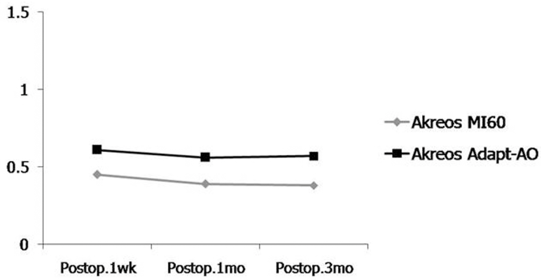
Figure 2. Changes of the surgically induced astigmatism in the two aspheric intraocular lens groups.
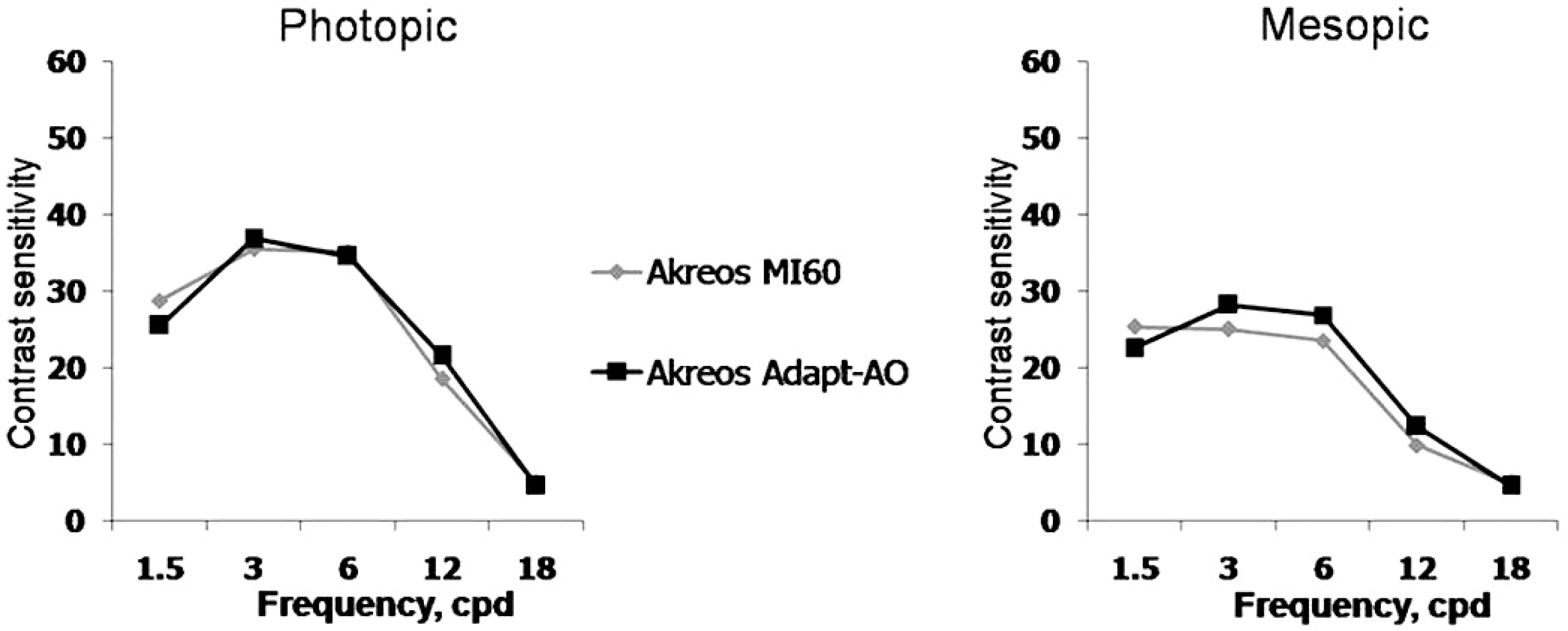
Figure 3. Comparison of contrast sensitivity test under photopic and mesopic condition at postoperative three months.
Reference
-
References
1. Kellman CD. abdominal and aspiration; a new technique of cataract removal; a preliminary report. Am J Ophthalmol. 1967; 64:23–35.2. Vasavada V, Vasavada V, Raj SM, Vasavada AR. Intraoperative abdominal and postoperative outcomes of microcoaxial phacoemulsification. Observational study. J Cataract Refract Surg. 2007; 33:1019–24.3. Yamaguchi T, Dogru M, Yamaguchi K, et al. Effect of spherical aberration on visual function under photopic and mesopic conditions after cataract surgery. J Cataract Refract Surg. 2009; 35:57–63.
Article4. Kershner RM. Clear corneal cataract surgery and the correction of myopia, hyperopia, and astigmatism. Ophthalmology. 1997; 104:381–9.
Article5. Cravy TV. Calculation of the change in corneal astigmatism following cataract extraction. Ophthalmic Surg. 1979; 10:38–49.6. Simsek S, Yasar T, Demirok A, et al. Effect of superior and temporal clear corneal incision on astigmatism after sutureless phacoemulsification. J Cataract Refract Surg. 1998; 24:515–8.7. Hwang SJ, Choi SK, Oh SH, et al. Surgically Induced Astigmatism and Corneal Higher Order Aberrations in Microcoaxial and Conventional Cataract Surgery. J Korean Ophthalmol Soc. 2008; 49:1597–602.
Article8. Werner L, Mamalis N. Wavefront corrections of intraocular lenses. Ophthalmol Clin North Am. 2004; 17:233–45.
Article9. Chalita MR, Krueger RR. Correlation of aberrations with visual acuity and symptoms. Ophthalmol Clin North Am. 2004; 17:135–42.
Article10. Guirao A, Redondo M, Geraghty E, et al. Corneal optical aberrations and retinal image quality in patients whom monofocal intraocular abdominal were implanted. Arch Ophthalmol. 2002; 120:1143–51.11. Wang L, Dai E, Koch DD, Nathoo A. Optical aberrations of the abdominal anterior cornea. J Cataract Refract Surg. 2003; 29:1514–21.12. Holladay JT, Piers PA, Koranyi G, et al. A new intraocular lens design to reduce spherical aberration of pseudophakic eyes. J Refract Surg. 2002; 18:683–91.
Article13. Dick HB. Recent developments in aspheric intraocular lenses. Curr Opin Ophthalmol. 2009; 20:25–32.
Article14. Altmann GE, Nichamin LD, Lane SS, Pepose JS. Optical performance of 3 intraocular lens designs in the presence of decentration. J Cataract Refract Surg. 2005; 31:574–85.
Article15. Holladay JT, Cravy TV, Koch DD. Calculating the surgically induced refractive change following ocular surgery. J Cataract Refract Surg. 1992; 18:429–43.
Article16. Huang FC, Teng SH. Compression of surgically induced astigmatism after sutureless temporal clear corneal and slceral frown incision. J Cataract Refract Surg. 1998; 24:477–81.17. Jee DH, Lee PY, Joo CK. The comparison of astigmatism according to the incision size in cataract operation. J Korean Ophthalmol Soc. 2003; 44:594–8.18. Kim HJ, Kim JH, Lee DH. Endothelial cell damage in microincision cataract surgery and coaxial phacoemulsification. J Korean Ophthalmol Soc. 2007; 48:19–26.19. Cavallini GM, Campi L, Masini C, et al. Bimanual microphacoemulsification vs coaxial miniphacoemulsification: Prospective study. J Cataract Refract Surg. 2007; 33:387–92.20. Choi JA, Chung SK, Kim HS. Comparative Study of Microcoaxial Cataract Surgery and Conventional Cataract Surgery. J Korean Ophthalmol Soc. 2008; 49:904–10.
Article21. Pandita D, Raj SM, Vasavada VA, et al. Contrast sensitivity and glare disability after implantation of acrySof IQ natural aspheric intraocular lens; prospective randomized masked clinical trial. J Cataract Refract Surg. 2007; 33:603–10.22. Oscher RH, Injev VP. Microcoaxial phacoemulsification Part 1: abdominal studies. J Cataract Refract Surg. 2007; 33:401–7.23. Oscher RH. Microcoaxial phacoemulsification Part 2: clinical studies. J cataract Refract Surg. 2007; 33:408–12.24. Alió J, Rodriguez-Prats JL, Galal A, Ramzy M. Outcomes of abdominal cataract versus coaxial phacoemulsification. Ophthalmology. 2005; 112:1997–2003.25. Masket S, Wang L, Belani S. Induced astigmatism with 2.2- and 3.0-mm coaxial phacoemulsification incisions. J Refract Surg. 2009; 25:21–4.
Article26. Hyashi K, Yoshida M, Hayashi H. Postoperative corneal shape changes: microincision versus small-incision coaxial cataract surgery. J Cataract Refract Surg. 2009; 35:233–9.27. Johansson B, Sundelin S, Wikberg-Matsson A, et al. Visual and optical performance of the Akreos Adapt Advanced Optics and Tecnis Z9000 intraocular lenses: Swedish multicenter study. J Cataract Refract Surg. 2007; 33:1565–72.28. Choi JA, Kim CY, Na KS, et al. Clinical results after implantation of a spherical aberration-free intraocular lens: Effect of contrast abdominal and wavefront aberration – a clinical comparative study. Ophthalmologica. 2009; 223:320–5.29. Shentu X, Tang X, Yao K. Spherical aberration, visual performance and pseudoaccommodation of eyes implanted with different aspheric intraocular lens. Clin Experiment Ophthalmol. 2008; 36:620–4.
Article
- Full Text Links
-
- Actions
-
Cited
- CITED
-
- Close
- Share
-
- Similar articles
-
- Comparison of Nd:YAG Capsulotomy Rates between Spherical and Aspheric Intraocular Lenses
- Comparison of the Clinical Effects of Implantation of Aspheric and Spherical Intraocular Lenses
- Clinical Efficacy of Bunny Multifocal Intraocular Lens after Cataract Surgery
- Comparative Study of Two Aspheric, Aberration-Free Intraocular Lenses in Cataract Surgery
- Spherical Aberration, Contrast Sensitivity and Depth of Focus With Three Aspherical Intraocular Lenses
