J Korean Ophthalmol Soc.
2009 Nov;50(11):1740-1744.
A Case of Isolated Choroidal Cystic Lesion With Good Visual Acuity
- Affiliations
-
- 1Department of Ophthalmology, Kang Dong Sacred Heart Hospital, Hallym Medical University, Seoul, Korea. sungpyo@hananet.net
- 2Department of Ophthalmology, Seoul National University College of Medicine, Seoul, Korea.
Abstract
- PURPOSE
To report a case of isolated choroidal cystic lesion in the macula with no interval change for two years.
CASE SUMMARY
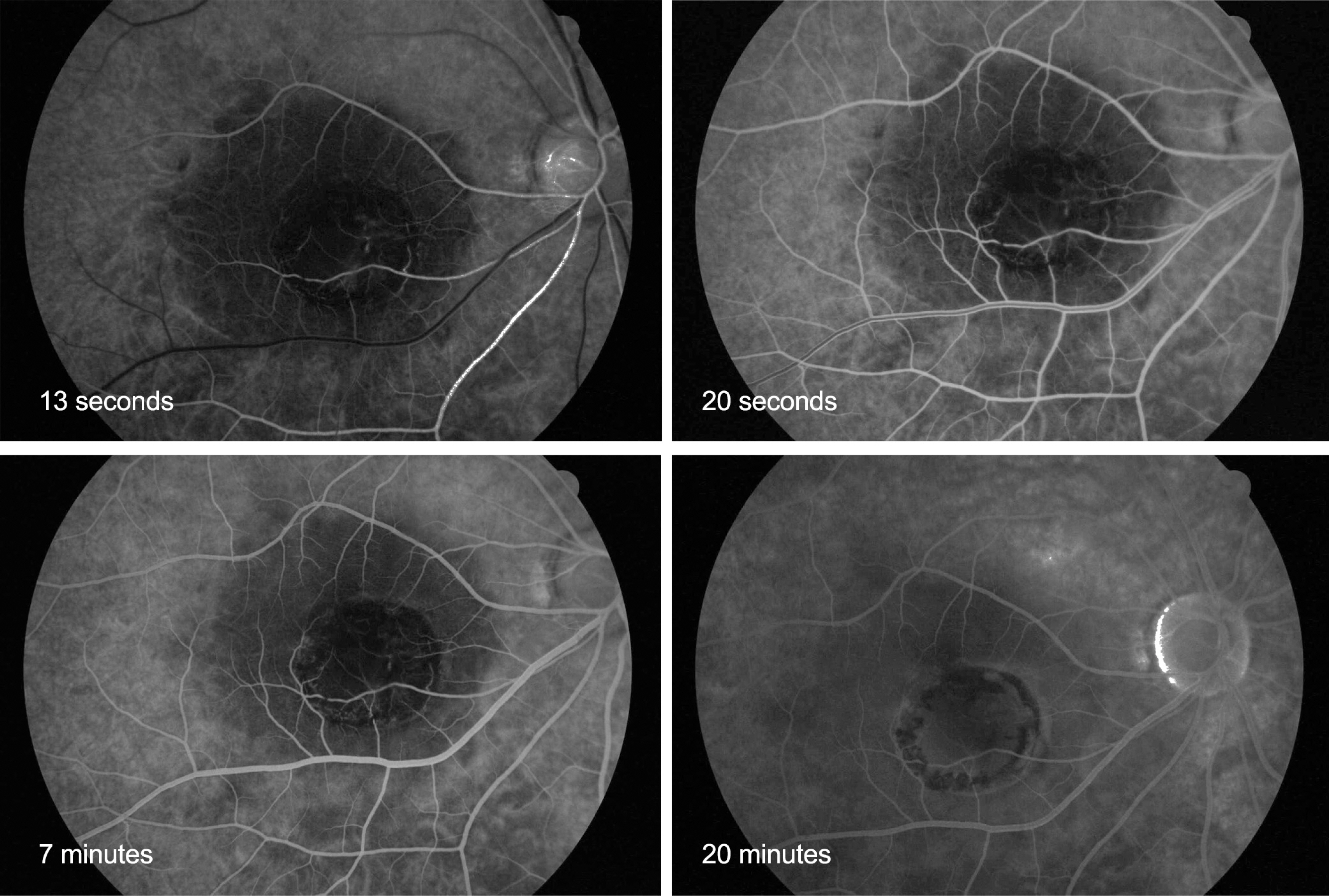
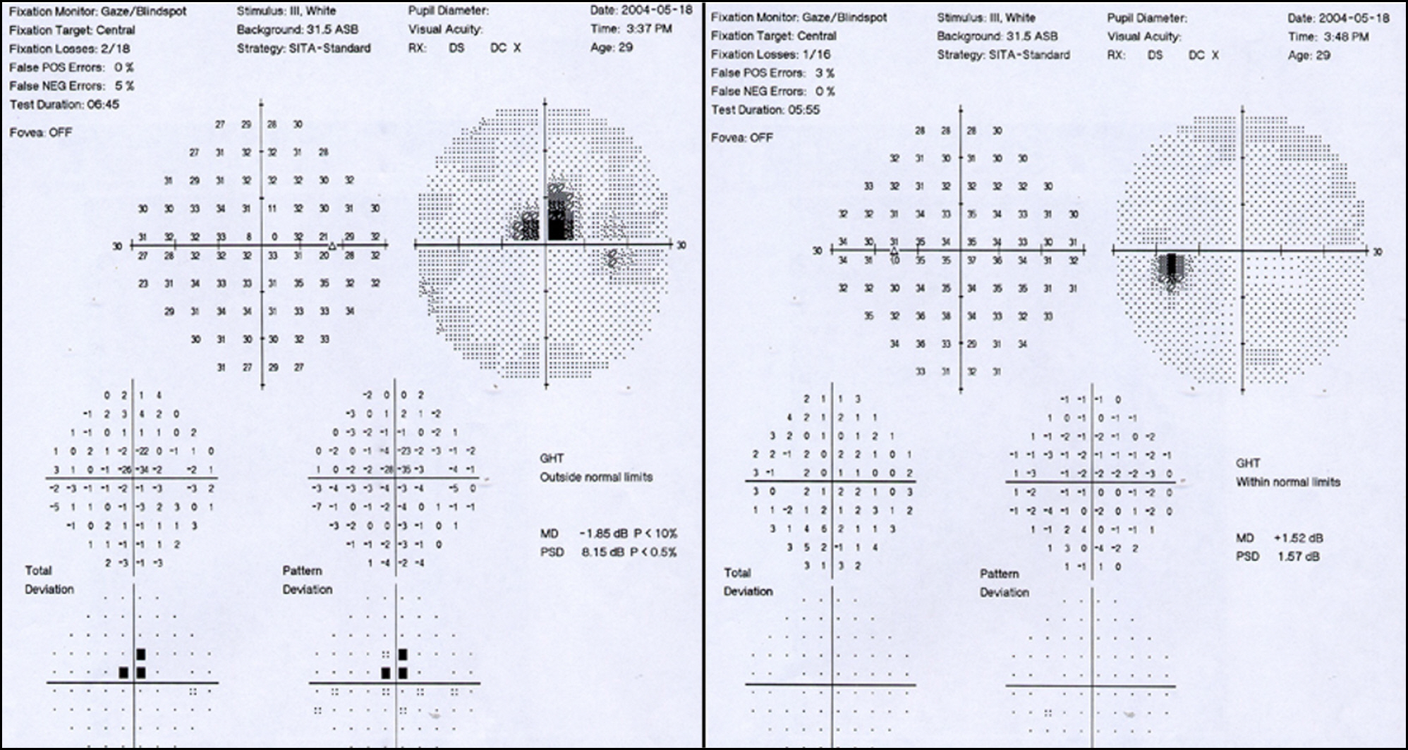
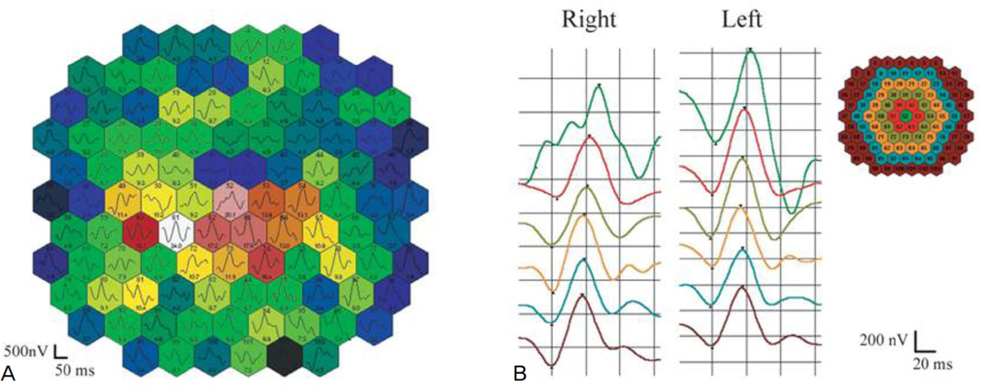
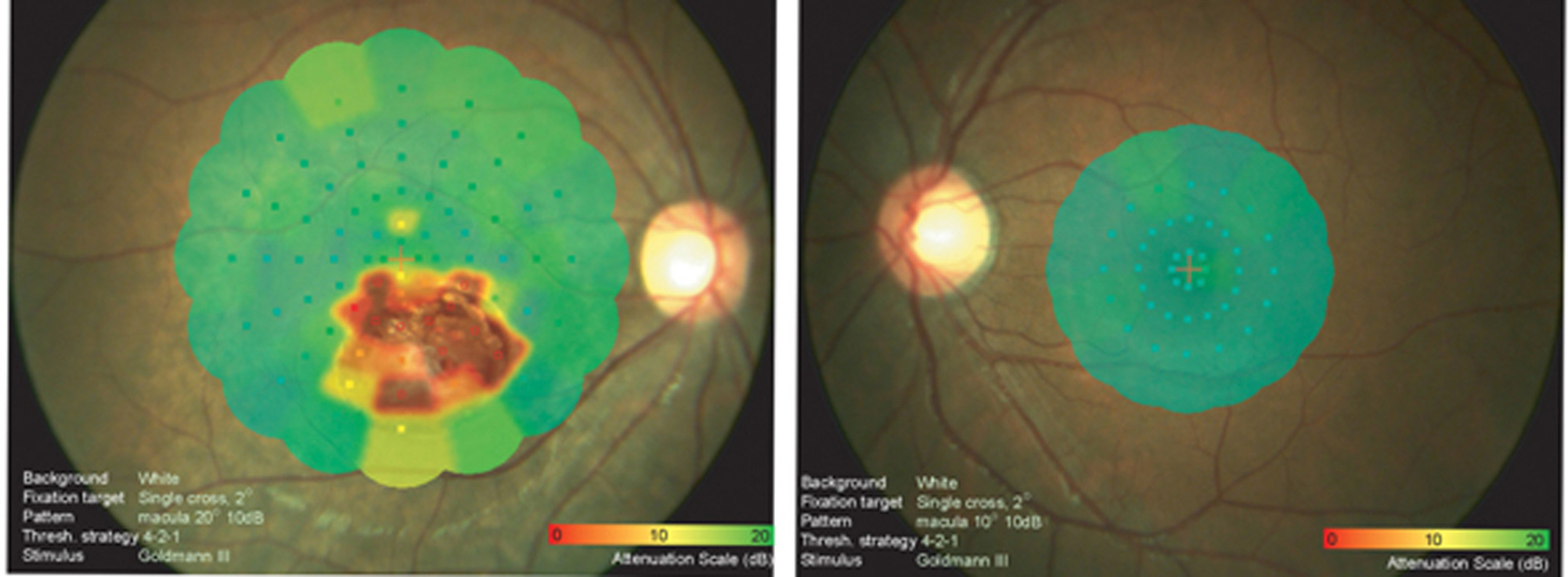
A 29-year-old woman who had suspicious maculopathy was referred to our clinic. Her best corrected visual acuity was 20/20 in the affected right eye, which showed a choroidal cystic lesion on a fundus exam, fluorescein angiography, USG and OCT. The multifocal ERG showed reduced amplitudes of the cystic area in the right eye, and SLO microperimetry revealed reduced retinal sensitivity in the cystic lesion as well as a stable fixation and spared foveal function. There was no evidence of underlying ocular disease in clinical assessment, and the lesion had not undergone interval change for the past two years.
Keyword
MeSH Terms
Figure
Reference
-
References
1. Espinoza G, Rosenblatt B, Harbour JW. Optical coherence tomography in the evaluation of retinal changes associated with suspicious choroidal melanocytic tumors. Am J Ophthalmol. 2004; 137:90–5.
Article2. Benhamou N, Massin P, Hauchine B, et al. Macular retinoschisis in highly myopic eyes. Am J Ophthalmol. 2002; 133:794–800.
Article3. Theodossiadis GP, Theodossiadis PG, Ladas ID, et al. Cyst formation in optic disc pit maculopathy. Doc Ophthalmol. 1999; 97:329–35.
Article4. Espinoza G, Rosenblatt B, Harbour JW. Optical coherence tomography in the evaluation of retinal changes associated with suspicious choroidal melanocytic tumors. Am J Ophthalmol. 2004; 137:90–5.
Article5. Higashide T, Akao N, Shirao E, Shirao Y. Optical coherence tomo-graphic and angiographic findings of a case with subretinal toxocara granuloma. Am J Ophthalmol. 2003; 136:188–90.
Article6. Shields CL, Honavar SG, Shiel JA, et al. Circumscribed choroidal hemangioma: clinical manifestations and factors predictive of visual outcome in 200 consecutive cases. Ophthalmology. 2001; 108:2237–48.
Article7. Fukushima T, Hirakawa T, Tanaka A, Tomonaga M. Choroidal epithelial cyst of the prepontine region: case report and ultrastructural study. Neurosurgery. 1988; 22:128–33.
Article
- Full Text Links
- Actions
-
Cited
- CITED
-
- Close
- Share
- Similar articles
-
- Choroidal Osteoma
- A Case of Choroidal Osteoma with Subretinal Hemorrhage Improved by Intravitreal Bevacizumab and Aflibercept Injections
- Photodynamic Therapy with Verteporfin in Polypoidal Choroidal Vasculopathy
- Effect of High-dose Intravitreal Bevacizumab Injection on Refractory Idiopathic Choroidal Neovasculariz
- A Case of Vogt-Koyanagi-Harada(VKH)Syndrome Combined with Choroidal Detachment in Diatetic Nephropathy Patient


