The Effect of Photodynamic Therapy in Chronic Central Serous Chorioretinopathy
- Affiliations
-
- 1Department of Ophthalmology, College of Medicine, The Catholic University of Korea, Seoul, Korea. wklee@catholic.ac.kr
Abstract
-
PURPOSE: To determine whether photodynamic therapy (PDT) is effective and safe for the treatment of chronic central serous chorioretinopathy (CSC) in Korean patients.
METHODS
Eleven eyes of 11 patients with chronic CSC underwent PDT. The laser spot size was chosen to cover the hyperfluorescent area on indocyanine green (ICG) angiography principally and to cover wide area including leaking point or detachment of retinal pigment epithelium on fluorescein angiography. We analyzed the medical records retrospectively to know the changes in visual acuity and serous detachment.
RESULTS
The mean follow-up was 14.2+/-6 months and exudative macular detachments resolved completely in 11 eyes within 1 month. In addition, eight weeks after treatment, the visual acuity improved in 3 eyes, and remained unchanged in 7 eyes and decreased in one eye. However, in 2 eyes, visual acuity decreased immediately after PDT due to choroidal hypoperfusion, and in one of them, the final visual acuity did not recover to the pre-treatment level.
CONCLUSIONS
In Korean patients, PDT seems to be effective for treatment of chronic CSC in the short term, but the long-term efficacy still must be determined. In addition, in PDT used for treatment of choroidal neovascularization (CNV), there was a possibility of irreversible occlusion in normal choroidal capillaries. Further studies are needed to determine the treatment parameters of PDT.
MeSH Terms
Figure
-

Figure 1. Fluorescein angiograph (FA) and optical coherence tomograph (OCT) of patient 4. Pre-treatment FA revealed leakage of dye under the sensory retina (A, B). OCT showed neurosensory retinal detachment as well as pigment epithelial detachment (PED) that was indistinct in FA (B). The visual acuity of this patient was 0.32 and photodynamic therapy (PDT) was done. One month after PDT, visual acuity improved to 0.63. FA revealed hyperfluorescence coming from retinal pigment epithelial window defect. Note the cessation of dye leakage (C, D). At this time, OCT showed complete resolution of PED and neurosensory retinal detachment (D).
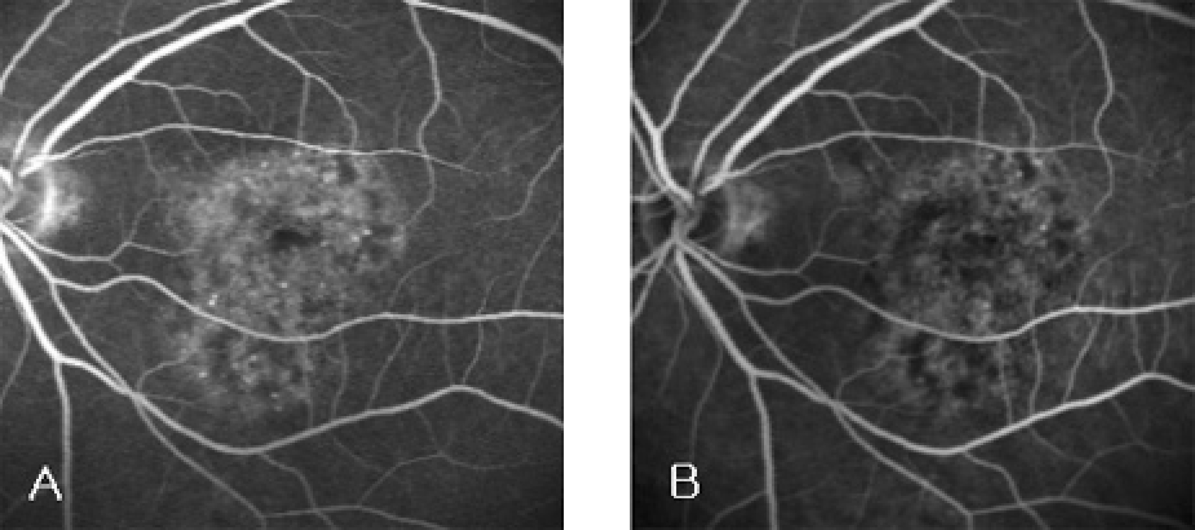
Figure 2. Fluorescein angiograph (FA) of patient 9. FA revealed diffuse leakage (A). The visual acuity in this patient was 0.4 and photodynamic therapy (PDT) was applied. Three months after PDT, the visual acuity improved to 0.5. In the FA taken at this time, no active leaking was observed (B).

Figure 3. Indocyanine green angiograph (ICGA) of patient 5. Pre-treatment ICGA showed diffuse choroidal hyperfluorescence in the macula (A). Three months after photodynamic therpy, ICGA showed decreased hyperfluorescence (B).
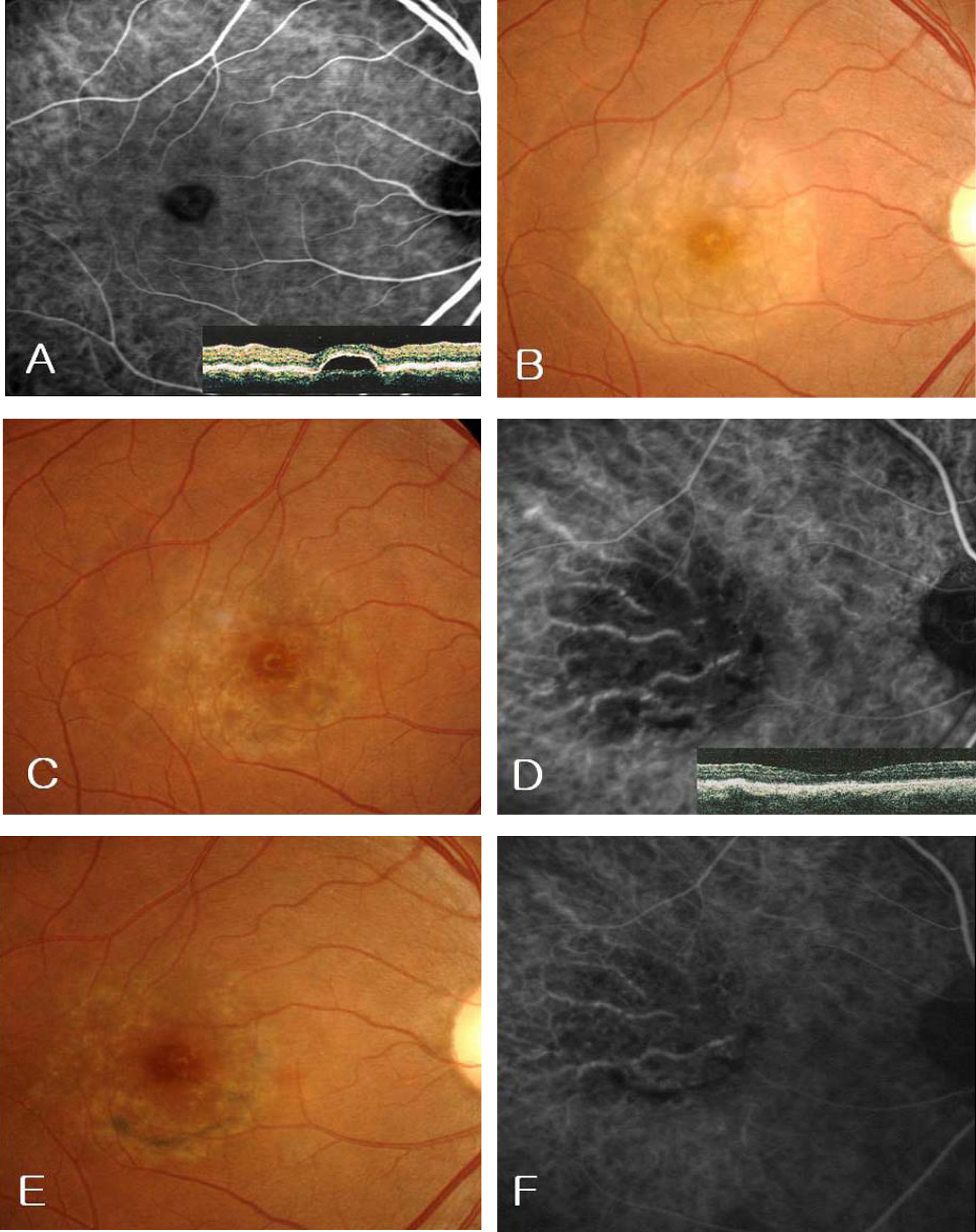
Figure 4. Choroidal ischemia after photodynamic therapy (PDT) in patient 3. Pre-treatment indocyanine green angiography (ICGA) and optical coherence tomography (OCT) showed retinal pigment epithelial detachment in the center of macula (A). In this patient, visual acuity was 1.0 and PDT was applied due to severe metamorphopsia. One week after PDT, visual acuity decreased to 0.3, and the fundus corresponding to the laser spot was pale (B). One month after PDT, visual acuity slightly improved to 0.6, but fundus photography showed still remained retinal whitening corresponding to the laser spot (C). At this time, pigment epithelial detachment was completely resolved in OCT but ICGA showed choroidal nonperfusion (D). One year after PDT, visual acuity improved to 1.0. At this time, fundus photography and ICGA revealed reperfusion of choriocapillary to some extent, but there was remained choroidal non-perfusion at the site of laser application (E, F).
Reference
-
References
1. Gass JD. Pathogenesis of disciform detachment of the neuro-epithelium. II. Idiopathic central serous choroidopathy. Am J Ophthalmol. 1967; 63:587–615.2. Gilbert CM, Owens SL, Smith PD, Fine SL. Long-term follow up of central serous choriortinopathy. Br J Ophthalmol. 1984; 68:815–20.3. Wang MS, Sander B, Larsen M. Retinal atrophy in idiopathic central serous chorioretinopathy. Am J Ophthalmol. 2002; 133:787–93.4. Idia T, Yannuzzi LA, Spaide RF, et al. Cystoid macular degeneration in chronic central serous chorioretinopathy. Retina. 2003; 23:1–7.
Article5. Yannuzzi LA. Laser photocoagulation of the macula: central serous chorioretinopathy. 1st ed.Philadelphia: JB Lippincott;1989. p. 1–12.6. Yanuzzi LA, Slakter JS, Kaufman SR, Gupta K. Laser treatment of diffuse retinal pigment epitheliopathy. Eur J Ophthalmol. 1992; 2:103–12.7. Robertson DM, Ilstrup D. Direct, indirect and sham laser photocoagulation in the management of central serous chorioretinopathy. Am J Ophthalmol. 1983; 95:457–66.
Article8. Guyer DR, Yannuzzi LA, Slakter JS, et al. Digital indocyaine green videoangiography of central serous chorioretinopathy. Arch Ophthalmol. 1994; 112:1057–62.9. Spaide RF, Hall L, Haas A, et al. Indocyanine green videoangiography of older patients with central serous chorioretinopathy. Retina. 1996; 16:203–13.
Article10. Bressler NM. Treatment of Age-related Macular Degeneration with Photodynamic Therapy (TAP) Study Group. Photodynamic therapy of subfoveal choroidal neovascularization in age-related macular degeneration with verteporfin: one-year results of 2 randomized clinical trials-TAP report 1. Arch Ophthalmol. 1999; 117:1329–45.11. Verteporfin in Photodynamic Therapy Study Group. Photodynamic therapy of subfoveal choroidal neovascularization in pathologic myopia with verteporfin: one-year results of a randomized clinical trial-VIP report 1. Ophthalmology. 2001; 108:841–52.12. Verteporfin in Photodynamic Therapy Study Group. Verteporfin therapy of subfoveal choroidal neovascularization in Age-related macular degeneration: two-year results of a randomized clinical trial including lesions with occult with no classic choroidal neovascularization-VIP report 2. Am J Ophthalmol. 2001; 131:541–60.13. Goshevsky JR, O'Day J. Photodynamic therapy in the management of juxtapapillary capillary hemangiomas. Clin Experiment Ophthalmol. 2005; 33:509–12.14. Yannuzzi LA, Slakter JS, Gross NE, et al. Indocyanine green angiography-guided photodynamic therapy for treatment of chronic central serous chorioretinopathy. Retina. 2003; 23:288–98.
Article15. Piccolino FC, Eandi CM, Ventre L, et al. Photodynamic therapy for chronic central serous chorioretinopathy. Retina. 2003; 23:752–63.
Article16. Taban M, Boyer DS, Thomas EL, Taban M. Chronic central serous chorioretinopathy: Photodynamic therapy. Am J Ophthalmol. 2004; 137:1073–80.
Article17. Chan WM, Lam DSC, Lai TYY, et al. Choroidal vascular remodelling in central serous chorioretinopathy after indocyanine green guided photodynamic therapy with verteporfin: a novel treatment at the primary disease level. Br J Ophthalmol. 2003; 87:1453–8.
Article18. Ober MD, Yannuzzi LA, Do DV, et al. Photodynamic therapy for focal retinal pigment epithelial leaks secondary to central serous chorioretinopathy. Ophthalmology. 2005; 112:2088–94.
Article19. Canakis C, Livir-Rallatos C, Panayiotis Z, et al. Ocular photodynamic therapy for serous macular detachment in the diffuse retinal pigment epitheliopathy variant of idiopathic central serous chorioretinopathy. Am J Ophthalmol. 2003; 136:750–2.
Article20. Olivier S, Harissi-Dagher M, Sebag M. Photodynamic therapy for chronic serous detachment of the retinal pigment epithelium in a young patient. Can J Ophthalmol. 2005; 40:214–6.
Article21. Lai TY, Chan WM, Li H, et al. Safety enhanced photodynamic therapy with half dose verteporfin for chronic central serous chorioretinopathy: a short term pilot study. Br J Ophthalmol. 2006; 90:869–74.
Article22. Schmidt-Erfurth U, Laqua H, Schlotzer-Schrehard U, et al. Histopathological changes following photodynamic therapy in human eyes. Arch Ophthalmol. 2002; 120:835–44.23. Michels S, Schmidt-Erfurth U. Sequence of early vascular events after photodynamic therapy. Invest Ophthalmol Vis Sci. 2003; 44:2147–54.
Article24. Schmidt-Erfurth U, Michels S, Barbazetto I, Laqua H. Photodynamic effects on choroidal neovascularization and physiological choroid. Invest Ophthalmol Vis Sci. 2002; 43:830–41.25. Prunte C, Flammer J. Choroidal capillary and venous congestion in central serous chorioretinopathy. Am J Ophthalmol. 1996; 121:26–34.26. Idia T, Kishi S, Hagimura N, Shimizu K. Persistent and bilateral choroidal vascular abnormalities in central serous chorioretinopathy. Retina. 1999; 19:508–12.
Article27. Scheider A, Nasemann JE, Lund OE. Fluorescein and indocyanine green angiographies of central serous choroidopathy by scanning laser ophthalmoscopy. Am J Ophthalmol. 1993; 115:50–6.
Article
- Full Text Links
-
- Actions
-
Cited
- CITED
-
- Close
- Share
-
- Similar articles
-
- Photodynamic Therapy With Verteporfin using Half Fluence for Chronic Central Serous Chorioretinopathy
- Review and update for central serous chorioretinopathy
- Photodynamic Therapy with Vertepofin for Short Time for Chronic Central Serous Chorioretinopathy
- A Case of Central Serous Chorioretinopathy Following Systemic Corticosteroid Therapy
- Electronmicroscopic Study of the Effect of Hexamethonium on Serous Choriretinopathy in Rabbits
