Distal Femoral Rotational Alignment Based on Mechanical Axis of the Femur: A 3-Dimensional Computed Tomographic Scan in Vivo Assessment
- Affiliations
-
- 1Department of Orthopedic Surgery, Konyang University College of Medicine, Daejeon, Korea. hurym1973@hanmail.net
- 2Department of Orthopedic Surgery, Yonsei University College of Medicine, Seoul, Korea.
Abstract
- PURPOSE
To measure and to analyze the relationships among the rotational axes of the distal femoral region by means of 3-dimensional (3D) computed tomographic (CT) images taken perpendicularly to the mechanical axis and a 3D rendering program.
MATERIALS AND METHODS
Fifty cases involving the lower extremity were included in this study, which used 3D computed tomographic angiograms. CT images of the perpendicular cross-sections to the mechanical axis of the femur were obtained by processing 3D recombinant images using Aquaris NET(R). The following anatomical angles were obtained from axial imaging using the 3D reconstructed bone model: transepicondylar axis, surgical transepicondylar axis, anteroposterior axis, and real posterior condylar axis.
RESULTS
In the tomographic images, the angles to the real posterior condylar axis formed by the anatomical femoral transepicondylar axis, the anatomical femoral transepicondylar axis, and the anteroposterior axis were 6.34+/-1.23degrees, 2.43+/-1.56degrees, and 96.52+/-1.77degrees, respectively. The angles to the anatomical femoral transepicondylar axis formed by the anteroposterior axis and the surgical femoral transepicondylar axis were 90.19+/-1.61degrees and 3.91+/-0.90degrees, respectively. In the recombinant 3D femur model, the angles to the real posterior condylar axis formed by the anatomical femoral transepicondylar axis and the anteroposterior axis were 6.29+/-1.86degrees, and 93.33+/-3.76degrees, respectively. And, the angle for anteroposterior axis from anatomical transepicondylar axis was 87.04+/-4.11degrees.
CONCLUSION
The method of measuring the rotational axes of the distal femur using the CT image taken perpendicularly to the mechanical axis is considered reproducible. In particular, the measurement method using the anatomical transepicondylar axis is more accurate than that using the anteroposterior axis.
Keyword
MeSH Terms
Figure
-
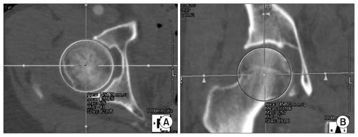
Figure 1 (A) The position of femoral head center (FHC) was determined by placing a circle onto the circumference of the femoral head on a set of axial and coronal section views, showing the largest bone contour of the femoral head. (B) The FHC locations were determined as the center of the circle that matched the largest bone contours.
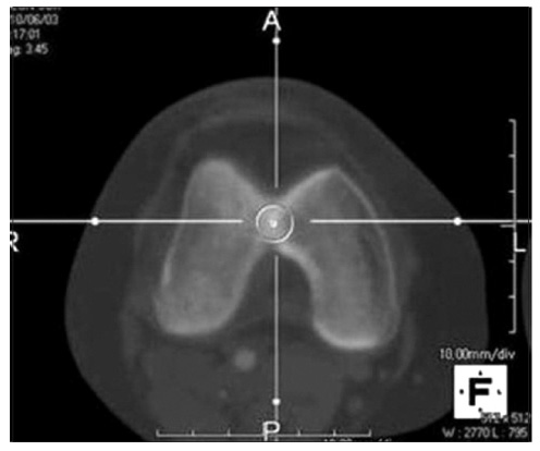
Figure 2 The intercondylar notch center was defined as the middle of the line connecting the narrowest anterior-to-posterior borders on an axial section of the distal femur.
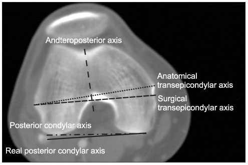
Figure 3 Construct of the reference axes on axial computed tomographic scan of the distal femoral condyle is shown.

Figure 4 Real posterior condylar axis was drawn. As the most lateral posterior condyle and the most medial posterior condyle did not exist on the same plane, real posterior condylar axis was drawn on the most medial posterior condylar plane by superimposing the most lateral posterior condyle to the medial plane. MAPC, medial apex of posterior condyle; LAPC, lateral apex of posterior condyle.

Figure 5 (A) Construct of the reference axes on 3-dimensional (3D) computed tomographic bone model of the distal femoral condyle is shown. 3D bone model was rotated until the center of femoral head was synchronized with the center of intercondylar notch. From caudocranial view, medial point of surgical transepicondylar axis was not detected because of hoarse-shoe shaped notch. (B) In obliquely rotated view, medial point of surgical transepicondylar axis is seen.
Reference
-
1. Victor J. Rotational alignment of the distal femur: a literature review. Orthop Traumatol Surg Res. 2009. 95:365–372.
Article2. Abadie P, Galaud B, Michaut M, Fallet L, Boisrenoult P, Beaufilsls P. Distal femur rotational alignment and patellar subluxation: a CT scan in vivo assessment. Orthop Traumatol Surg Res. 2009. 95:267–271.
Article3. Newbern DG, Faris PM, Ritter MA, Keating EM, Meding JB, Berend ME. A clinical comparison of patellar tracking using the transepicondylar axis and the posterior condylar axis. J Arthroplasty. 2006. 21:1141–1146.
Article4. Walde TA, Bussert J, Sehmisch S, et al. Optimized functional femoral rotation in navigated total knee arthroplasty considering ligament tension. Knee. 2010. 17:381–386.
Article5. Berger RA, Crossett LS, Jacobs JJ, Rubash HE. Malrotation causing patellofemoral complications after total knee arthroplasty. Clin Orthop Relat Res. 1998. (356):144–153.
Article6. Kingsley PC, Olmsted KL. A study to determine the angle of anteversion of the neck of the femur. J Bone Joint Surg Am. 1948. 30A:745–751.
Article7. Lee YS, Oh SH, Seon JK, Song EK, Yoon TR. 3D femoral neck anteversion measurements based on the posterior femoral plane in ORTHODOC system. Med Biol Eng Comput. 2006. 44:895–906.
Article8. Kim JS, Park TS, Park SB, Kim JS, Kim IY, Kim SI. Measurement of femoral neck anteversion in 3D. Part 1: 3D imaging method. Med Biol Eng Comput. 2000. 38:603–609.
Article9. Griffin FM, Insall JN, Scuderi GR. The posterior condylar angle in osteoarthritic knees. J Arthroplasty. 1998. 13:812–815.
Article10. Victor J, Van Doninck D, Labey L, Van Glabbeek F, Parizel P, Bellemans J. A common reference frame for describing rotation of the distal femur: a ct-based kinematic study using cadavers. J Bone Joint Surg Br. 2009. 91:683–690.11. Yau WP, Leung A, Liu KG, Yan CH, Wong LL, Chiu KY. Interobserver and intra-observer errors in obtaining visually selected anatomical landmarks during registration process in non-image-based navigation-assisted total knee arthroplasty. J Arthroplasty. 2007. 22:1150–1161.
Article12. Siston RA, Patel JJ, Goodman SB, Delp SL, Giori NJ. The variability of femoral rotational alignment in total knee arthroplasty. J Bone Joint Surg Am. 2005. 87:2276–2280.
Article13. Jenny JY, Boeri C. Low reproducibility of the intra-operative measurement of the transepicondylar axis during total knee replacement. Acta Orthop Scand. 2004. 75:74–77.
Article14. Stoeckl B, Nogler M, Krismer M, Beimel C, de la Barrera JL, Kessler O. Reliability of the transepicondylar axis as an anatomical landmark in total knee arthroplasty. J Arthroplasty. 2006. 21:878–882.
Article15. Arima J, Whiteside LA, McCarthy DS, White SE. Femoral rotational alignment, based on the anteroposterior axis, in total knee arthroplasty in a valgus knee. A technical note. J Bone Joint Surg Am. 1995. 77:1331–1334.
Article16. Yoshioka Y, Siu D, Cooke TD. The anatomy and functional axes of the femur. J Bone Joint Surg Am. 1987. 69:873–880.
Article17. Servien E, Viskontas D, Giuffrè BM, Coolican MR, Parker DA. Reliability of bony landmarks for restoration of the joint line in revision knee arthroplasty. Knee Surg Sports Traumatol Arthrosc. 2008. 16:263–269.
Article18. Stiehl JB, Abbott BD. Morphology of the transepicondylar axis and its application in primary and revision total knee arthroplasty. J Arthroplasty. 1995. 10:785–789.
Article19. Berger RA, Rubash HE, Seel MJ, Thompson WH, Crossett LS. Determining the rotational alignment of the femoral component in total knee arthroplasty using the epicondylar axis. Clin Orthop Relat Res. 1993. (286):40–47.
Article20. Griffin FM, Math K, Scuderi GR, Insall JN, Poilvache PL. Anatomy of the epicondyles of the distal femur: MRI analysis of normal knees. J Arthroplasty. 2000. 15:354–359.21. Nagamine R, Miura H, Inoue Y, et al. Reliability of the anteroposterior axis and the posterior condylar axis for determining rotational alignment of the femoral component in total knee arthroplasty. J Orthop Sci. 1998. 3:194–198.22. Asano T, Akagi M, Nakamura T. The functional flexion-extension axis of the knee corresponds to the surgical epicondylar axis: in vivo analysis using a biplanar image-matching technique. J Arthroplasty. 2005. 20:1060–1067.23. Yoshino N, Takai S, Ohtsuki Y, Hirasawa Y. Computed tomography measurement of the surgical and clinical transepicondylar axis of the distal femur in osteoarthritic knees. J Arthroplasty. 2001. 16:493–497.
Article24. Moon YW, Seo JG, Lim SJ, Yang JH. Variability in femoral component rotation reference axes measured during navigation-assisted total knee arthroplasty using gap technique. J Arthroplasty. 2010. 25:238–243.
Article25. Matsuda S, Miura H, Nagamine R, et al. Anatomical analysis of the femoral condyle in normal and osteoarthritic knees. J Orthop Res. 2004. 22:104–109.
Article
- Full Text Links
-
- Actions
-
Cited
- CITED
-
- Close
- Share
-
- Similar articles
-
- Measurement of the Axial Rotational Axis of Distal Femur Using Different Landmarks
- Anatomical Assessment of the Distal Femur and Tibia for Optimal Femoral Rotational Alignment in Total Knee Arthroplasty
- Anatomical Assessment of Distal Femur for Optimal Femoral Component Rotational Alignment in TKA
- Bony Landmarks for Determining the Mechanical Axis of the Femur in the Sagittal Plane during Total Knee Arthroplasty
- MRI Measurement of the Distal Femur for Determining the Rotational Alignment in TKA
