Korean J Clin Neurophysiol.
2016 Jun;18(1):18-20. 10.14253/kjcn.2016.18.1.18.
Myelin Water Fraction MRI in a Case of Clinically Probable Amyotrophic Lateral Sclerosis
- Affiliations
-
- 1Department of Neurology, Gachon University Gil Medical Center, Incheon, Korea. dr.donghoon.shin@gmail.com
- 2Laboratory for Imaging Science and Technology (LIST), Department of Electrical and Computer Engineering, Seoul National University, Seoul, Korea.
- 3Department of Radiology, Gachon University Gil Medical Center, Incheon, Korea.
- KMID: 2328824
- DOI: http://doi.org/10.14253/kjcn.2016.18.1.18
Abstract
- Amyotrophic lateral sclerosis (ALS) is a progressive motor neuron degenerative disease that clinically manifests both upper and lower motor neuron signs. However, it is unknown where and how the motor neuron degeneration begins, and conflicting hypotheses have been suggested. Recent advanced radiological techniques enable us to look into ALS neuropathology in vivo. Herein, we report a case with upper motor neuron-predominant ALS in whom the results of brain magnetic resonance imaging (MRI) and myelin water fraction MRI suggest axonal degeneration.
MeSH Terms
Figure
-
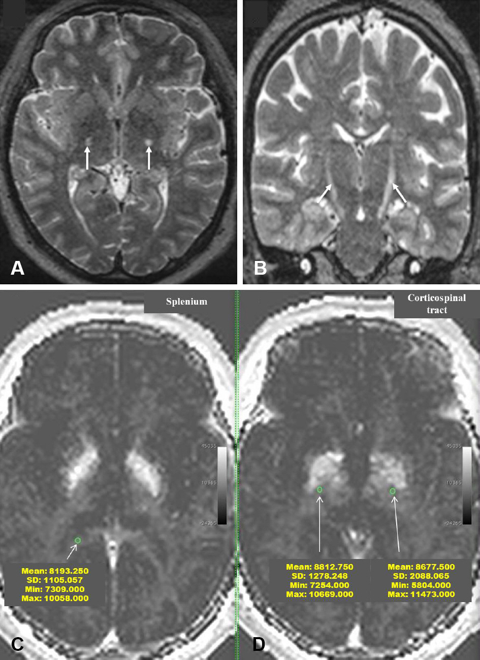
Figure 1. (A, B) Axial (A) and coronal (B) T2-weighted MRI of the brain display bilateral high signal intensity along the corticospinal tract (CST) (white arrows). (C, D) The mean myelin quantity was larger in the CST area than in the adjacent splenium of corpus callosum (right CST = 8,812, left CST = 8,677, splenium = 8,193) (green circles). MRI; magnetic resonance image.
Reference
-
1.Kiernan MC., Vucic S., Cheah BC., Turner MR., Eisen A., Hardiman O, et al. Amyotrophic lateral sclerosis. Lancet. 2011. 377:942–955.
Article2.Brooks BR., Miller RG., Swash M., Munsat TL. World Federation of Neurology Research Group on Motor Neuron Diseases. El Escorial revisited: revised criteria for the diagnosis of amyotrophic lateral sclerosis. Amyotroph Lateral Scler Other Motor Neuron Disord. 2000. 1:293–299.
Article3.Turner MR., Agosta F., Bede P., Govind V., Lule D., Verstraete E. Neuroimaging in amyotrophic lateral sclerosis. Biomark Med. 2012. 6:319–337.
Article4.Zhang L., Ulug AM., Zimmerman RD., Lin MT., Rubin M., Beal MF. The diagnostic utility of FLAIR imaging in clinically verified amyotrophic lateral sclerosis. J Magn Reson Imaging. 2003. 17:521–527.
Article5.Mirowitz S., Sartor K., Gado M., Torack R. Focal signal-intensity variations in the posterior internal capsule: normal MR findings and distinction from pathologic findings. Radiology. 1989. 172:535–539.
Article6.Córdoba J., Raguer N., Flavià M., Vargas V., Jacas C., Alonso J, et al. T2 hyperintensity along the cortico-spinal tract in cirrhosis relates to functional abnormalities. Hepatology. 2003. 38:1026–1033.
Article7.Waragai M. MRI and clinical features in amyotrophic lateral sclerosis. Neuroradiology. 1997. 39:847–851.
Article8.Vucic S., Ziemann U., Eisen A., Hallett M., Kiernan MC. Transcranial magnetic stimulation and amyotrophic lateral sclerosis: pathophysiological insights. J Neurol Neurosurg Psychiatry. 2013. 84:1161–1170.
Article9.Borich MR., Mackay AL., Vavasour IM., Rauscher A., Boyd LA. Evaluation of white matter myelin water fraction in chronic stroke. Neuroimage Clin. 2013. 2:569–580.
Article10.Kolind S., Sharma R., Knight S., Johansen-Berg H., Talbot K., Turner MR. Myelin imaging in amyotrophic and primary lateral sclerosis. Amyotroph Lateral Scler Frontotemporal Degener. 2013. 14:562–573.
Article
- Full Text Links
-
- Actions
-
Cited
- CITED
-
- Close
- Share
-
- Similar articles
-
- Syndrome of Progressive Bulbar Palsy in Amyotrophic Lateral Sclerosis: A Case Report
- Amyotrophic Lateral Sclerosis Associated With CADASIL
- Apraxia of Eyelid Closure and Motor Impersistence of Eyelid in a Patient with Amyotrophic Lateral Sclerosis
- A Case of Frontotemporal Dementia with Amyotrophic Lateral Sclerosis Presenting with Pathological Gambling
- Psychosocial Responses and Quality of Life among Amyotrophic Lateral Sclerosis Patients and Their Caregivers
