J Korean Assoc Oral Maxillofac Surg.
2014 Jun;40(3):103-110.
Evaluation of soft tissue asymmetry using cone-beam computed tomography after open reduction and internal fixation of zygomaticomaxillary complex fracture
- Affiliations
-
- 1Department of Oral and Maxillofacial Surgery, College of Dentistry, Wonkwang University, Iksan, Korea. kkhoms@hanmail.net
- 2Wonkwang Dental Research Institute, Wonkwang University, Iksan, Korea.
- 3Department of Oral and Maxillofacial Surgery, Graduate School of Dentistry, Wonkwang University, Iksan, Korea.
Abstract
OBJECTIVES
In this study, we assessed soft tissue asymmetry that occurred after open reduction of unilateral zygomaticomaxillary complex (ZMC) fractures. We proposed a simple method to assess soft tissue asymmetry after reduction surgery by evaluating the symmetry between the affected and the unaffected sides. The factors affecting soft tissue contour after surgery were also analyzed.
MATERIALS AND METHODS
Subjects included patients admitted to Wonkwang University Dental Hospital from 2008 to 2013. Cone-beam computed tomography (CBCT) images of asymmetric patients who underwent open reduction at least 3 months prior were compared with healthy patients.
RESULTS
The degree of asymmetry was measured in both the open reduction and control groups. Landmarks that showed a statistically significant difference between the two groups were zygion (1.73+/-0.24 mm), bucclae (1.08+/-0.26 mm), point of cheek (2.05+/-0.33 mm) and frontozygomatic point (1.30+/-0.31 mm).
CONCLUSION
When compared with the normal group, asymmetry can occur in the affected side, which usually shows depression of overlying soft tissue and is statistically significantly different. Evaluation of soft tissue asymmetry with CBCT images after open reduction of ZMC fracture is useful.
Keyword
Figure
-
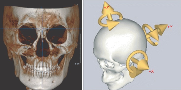
Fig. 1 Constructed coordinate system used. X-axis (transverse axis) is a line parallel to the frontozygomatic (FZ) line. Y-axis (anteroposterior axis) is a line perpendicular to FZ line while parallel to right Frankfort horizontal (R FH) plane. Z-axis is perpendicular to both FZ line and R FH plane.
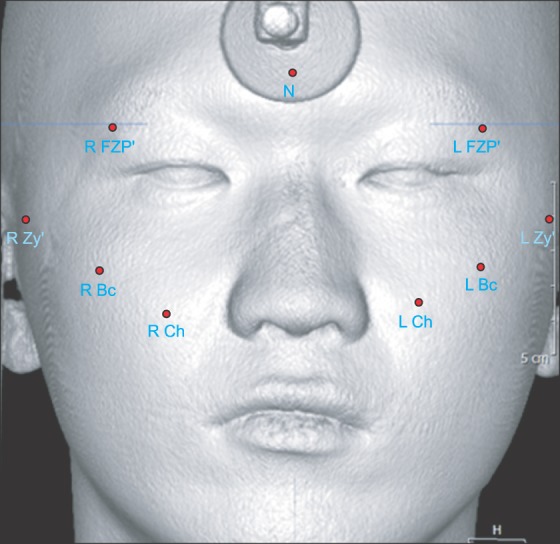
Fig. 2 Soft tissue landmarks, nasion (N), zygion' (Zy'), buccale (Bc), point of cheek (Ch), and frontozygomatic point' (FZP'). (R: right, L: left)
Reference
-
1. Ellis E 3rd, Kittidumkerng W. Analysis of treatment for isolated zygomaticomaxillary complex fractures. J Oral Maxillofac Surg. 1996; 54:386–400. PMID: 8600255.
Article2. Chae YP, Kim SK. Clinical study on surgical treatment of zygoma fractures. J Korean Dent Assoc. 1989; 27:949–957.3. Toriumi M, Nagasao T, Itamiya T, Shimizu Y, Yasudo H, Sakamoto Y, et al. 3-D analysis of dislocation in zygoma fractures. J Craniomaxillofac Surg. 2013; doi: 10.1016/j.jcms.2013.06.003.
Article4. Czerwinski M, Ma S, Williams HB. Zygomatic arch deformation: an anatomic and clinical study. J Oral Maxillofac Surg. 2008; 66:2322–2329. PMID: 18940500.
Article5. Zhang QB, Dong YJ, Guan JB, Li ZB, Zhao JH, Dong FS. Epidemiology and treatment of fractures of the zygomatic complex. Asian J Oral Maxillofac Surg. 2008; 20:59–64.
Article6. Ellis E 3rd, el-Attar A, Moos KF. An analysis of 2,067 cases of zygomatico-orbital fracture. J Oral Maxillofac Surg. 1985; 43:417–428. PMID: 3858478.
Article7. Gaziri DA, Omizollo G, Luchi GH, de Oliveira MG, Heitz C. Assessment for treatment of tripod fractures of the zygoma with microcompressive screws. J Oral Maxillofac Surg. 2012; 70:e378–e388. PMID: 22608820.
Article8. Miller L, Morris DO, Berry E. Visualizing three-dimensional facial soft tissue changes following orthognathic surgery. Eur J Orthod. 2007; 29:14–20. PMID: 16957060.
Article9. Solem RC, Marasco R, Guiterrez-Pulido L, Nielsen I, Kim SH, Nelson G. Three-dimensional soft-tissue and hard-tissue changes in the treatment of bimaxillary protrusion. Am J Orthod Dentofacial Orthop. 2013; 144:218–228. PMID: 23910203.
Article10. Baik HS, Jeon JM, Lee HJ. Facial soft-tissue analysis of Korean adults with normal occlusion using a 3-dimensional laser scanner. Am J Orthod Dentofacial Orthop. 2007; 131:759–766. PMID: 17561054.
Article11. Li H, Yang Y, Chen Y, Wu Y, Zhang Y, Wu D, et al. Three-dimensional reconstruction of maxillae using spiral computed tomography and its application in postoperative adult patients with unilateral complete cleft lip and palate. J Oral Maxillofac Surg. 2011; 69:e549–e557. PMID: 21982692.
Article12. Damstra J, Oosterkamp BC, Jansma J, Ren Y. Combined 3-dimensional and mirror-image analysis for the diagnosis of asymmetry. Am J Orthod Dentofacial Orthop. 2011; 140:886–894. PMID: 22133955.
Article13. Cavalcanti MG, Rocha SS, Vannier MW. Craniofacial measurements based on 3D-CT volume rendering: implications for clinical applications. Dentomaxillofac Radiol. 2004; 33:170–176. PMID: 15371317.
Article14. Park SH, Yu HS, Kim KD, Lee KJ, Baik HS. A proposal for a new analysis of craniofacial morphology by 3-dimensional computed tomography. Am J Orthod Dentofacial Orthop. 2006; 129:600.e23–600.e34. PMID: 16679198.
Article15. You KH, Lee KJ, Lee SH, Baik HS. Three-dimensional computed tomography analysis of mandibular morphology in patients with facial asymmetry and mandibular prognathism. Am J Orthod Dentofacial Orthop. 2010; 138:540.e1–540.e8. PMID: 21055584.
Article16. Lee MS, Chung DH, Lee JW, Cha KS. Assessing soft-tissue characteristics of facial asymmetry with photographs. Am J Orthod Dentofacial Orthop. 2010; 138:23–31. PMID: 20620830.
Article17. Baik HS, Kim SY. Facial soft-tissue changes in skeletal class III orthognathic surgery patients analyzed with 3-dimensional laser scanning. Am J Orthod Dentofacial Orthop. 2010; 138:167–178. PMID: 20691358.
Article18. Meyer-Marcotty P, Alpers GW, Gerdes AB, Stellzig-Eisenhauer A. Impact of facial asymmetry in visual perception: a 3-dimensional data analysis. Am J Orthod Dentofacial Orthop. 2010; 137:168.e1–168.e8. PMID: 20152669.
Article19. Peck S, Peck L, Kataja M. Skeletal asymmetry in esthetically pleasing faces. Angle Orthod. 1991; 61:43–48. PMID: 2012321.20. Ferrario VF, Sforza C, Poggio CE, Serrao G. Facial three-dimensional morphometry. Am J Orthod Dentofacial Orthop. 1996; 109:86–93. PMID: 8540488.
Article21. Furst IM, Austin P, Pharoah M, Mahoney J. The use of computed tomography to define zygomatic complex position. J Oral Maxillofac Surg. 2001; 59:647–654. PMID: 11381388.
Article22. Gwilliam JR, Cunningham SJ, Hutton T. Reproducibility of soft tissue landmarks on three-dimensional facial scans. Eur J Orthod. 2006; 28:408–415. PMID: 16901962.
Article23. Cho HJ. A three-dimensional cephalometric analysis. J Clin Orthod. 2009; 43:235–252. PMID: 19458456.24. Modabber A, Rana M, Ghassemi A, Gerressen M, Gellrich NC, Hölzle F, et al. Three-dimensional evaluation of postoperative swelling in treatment of zygomatic bone fractures using two different cooling therapy methods: a randomized, observer-blind, prospective study. Trials. 2013; 14:238. PMID: 23895539.
Article25. Hwang HS, Yuan D, Jeong KH, Uhm GS, Cho JH, Yoon SJ. Three-dimensional soft tissue analysis for the evaluation of facial asymmetry in normal occlusion individuals. Korean J Orthod. 2012; 42:56–63. PMID: 23112933.
Article26. Zachariades N, Mezitis M, Anagnostopoulos D. Changing trends in the treatment of zygomaticomaxillary complex fractures: a 12-year evaluation of methods used. J Oral Maxillofac Surg. 1998; 56:1152–1156. PMID: 9766540.
Article27. Kubota Y, Kuroki T, Akita S, Koizumi T, Hasegawa M, Rikihisa N, et al. Association between plate location and plate removal following facial fracture repair. J Plast Reconstr Aesthet Surg. 2012; 65:372–378. PMID: 22030077.
Article28. Klotch DW, Gilliland R. Internal fixation vs. conventional therapy in midface fractures. J Trauma. 1987; 27:1136–1145. PMID: 3312622.
Article29. Alpert B, Tiwana PS, Kushner GM. Management of comminuted fractures of the mandible. Oral Maxillofac Surg Clin North Am. 2009; 21:185–192. PMID: 19348983.
Article30. Ferrario VF, Sforza C, Schmitz JH, Miani A Jr, Serrao G. A three-dimensional computerized mesh diagram analysis and its application in soft tissue facial morphometry. Am J Orthod Dentofacial Orthop. 1998; 114:404–413. PMID: 9790324.
Article31. af Geijerstam B, Hultman G, Bergström J, Stjärne P. Zygomatic fractures managed by closed reduction: an analysis with postoperative computed tomography follow-up evaluating the degree of reduction and remaining dislocation. J Oral Maxillofac Surg. 2008; 66:2302–2307. PMID: 18940496.
Article32. Wittwer G, Adeyemo WL, Yerit K, Voracek M, Turhani D, Watzinger F, et al. Complications after zygoma fracture fixation: is there a difference between biodegradable materials and how do they compare with titanium osteosynthesis? Oral Surg Oral Med Oral Pathol Oral Radiol Endod. 2006; 101:419–425. PMID: 16545702.
Article33. Langford RJ, Frame JW. Tissue changes adjacent to titanium plates in patients. J Craniomaxillofac Surg. 2002; 30:103–107. PMID: 12069513.
Article34. Dal Santo F, Ellis E 3rd, Throckmorton GS. The effects of zygomatic complex fracture on masseteric muscle force. J Oral Maxillofac Surg. 1992; 50:791–799. PMID: 1634969.
Article
- Full Text Links
-
- Actions
-
Cited
- CITED
-
- Close
- Share
-
- Similar articles
-
- Corrigendum: Evaluation of soft tissue asymmetry using cone-beam computed tomography after open reduction and internal fixation of zygomaticomaxillary complex fracture
- Clinical Experiences of Facial Asymmetries in Zygomaticomaxillary Complex Bone Fracture Patients
- Differences in facial soft tissue deviations in Class III patients with different types of mandibular asymmetry: A cone-beam computed tomography study
- Zygomaticomaxillary complex fracture after two-jaw surgery
- Retrospective study about the postoperative stability of zygomaticomaxillary complex fracture
