Investig Magn Reson Imaging.
2016 Jun;20(2):81-87. 10.13104/imri.2016.20.2.81.
Detecting Peripheral Nerves in the Elbow using Three-Dimensional Diffusion-Weighted PSIF Sequences: a Feasibility Pilot Study
- Affiliations
-
- 1Department of Radiology, Korea University Guro Hospital, Korea University College of Medicine, Seoul, Korea. hongsj@korea.ac.kr
- 2Department of Radiology, Sejong General Hospital, Gyeonggi-do, Korea.
- 3Department of Radiology, Korea University Anam Hospital, Korea University College of Medicine, Seoul, Korea.
- 4Department of Radiology, Korea University Ansan Hospital, Korea University College of Medicine, Gyeonggi-do, Korea.
- KMID: 2327419
- DOI: http://doi.org/10.13104/imri.2016.20.2.81
Abstract
- PURPOSE
To analyze the feasibility of three-dimensional (3D) diffusion-weighted (DW) PSIF (reversed FISP [fast imaging with steady-state free precession]) sequence in order to evaluate peripheral nerves in the elbow.
MATERIALS AND METHODS
Ten normal, asymptomatic volunteers were enrolled (6 men, 4 women, mean age 27.9 years). The following sequences of magnetic resonance images (MRI) of the elbow were obtained using a 3.0-T machine: 3D DW PSIF, 3D T2 SPACE (sampling perfection with application optimized contrasts using different flip angle evolution) with SPAIR (spectral adiabatic inversion recovery) and 2D T2 TSE (turbo spin echo) with modified Dixon (m-Dixon) sequence. Two observers used a 5-point grading system to analyze the image quality of the ulnar, median, and radial nerves. The signal-to-noise ratio (SNR) and contrast-to-noise ratio (CNR) of each nerve were measured. We compared 3D DW PSIF images with other sequences using the Wilcoxon-signed rank test and Friedman test. Inter-observer agreement was measured using intraclass correlation coefficient (ICC) analysis.
RESULTS
The mean 5-point scores of radial, median, and ulnar nerves in 3D DW PSIF (3.9/4.2/4.5, respectively) were higher than those in 3D T2 SPACE SPAIR (1.9/2.8/2.8) and 2D T2 TSE m-Dixon (1.7/2.8/2.9) sequences (P < 0.05). The mean SNR in 3D DW PSIF was lower than 3D T2 SPACE SPAIR, but there was no difference between 3D DW PSIF and 2D T2 TSE m-Dixon in all of the three nerves. The mean CNR in 3D DW PSIF was lower than 3D T2 SPACE SPAIR and 2D T2 TSE m-Dixon in the median and ulnar nerves, but no difference among the three sequences in the radial nerve.
CONCLUSION
The three-dimensional DW PSIF sequence may be feasible to evaluate the peripheral nerves around the elbow in MR imaging. However, further optimization of the image quality (SNR, CNR) is required.
Keyword
MeSH Terms
Figure
-

Fig. 1 Qualitative analysis score of each nerve.

Fig. 2 MR neurography images. (a) Fat-suppressed 3D DW PSIF image, (b) Fat-suppressed 3D T2 SPAIR image, (c) Fatsuppressed 2D T2 m-Dixon image. In the fat-suppressed 3D DWI PSIF images, the median (thick arrows), radial (arrows) and ulnar nerves (curved arrows) are easily defined with suppressed adjacent vessel signals. However, in other sequences, it is difficult to differentiate the nerves from the adjacent vessel signals, even though the SNR and CNR of the nerves are better than are the fat-suppressed 3D DW PSIF images.
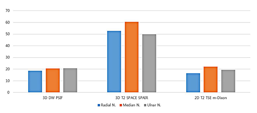
Fig. 3 Quantitative analysis score of each nerve (SNR).

Fig. 4 Quantitative analysis score of each nerve (CNR).
Reference
-
1. Chhabra A, Andreisek G, Soldatos T, et al. MR neurography: past, present, and future. AJR Am J Roentgenol. 2011; 197:583–591.2. Thawait SK, Chaudhry V, Thawait GK, et al. High-resolution MR neurography of diffuse peripheral nerve lesions. AJNR Am J Neuroradiol. 2011; 32:1365–1372.3. Chhabra A, Flammang A, Padua A Jr, Carrino JA, Andreisek G. Magnetic resonance neurography: technical considerations. Neuroimaging Clin N Am. 2014; 24:67–78.4. Chhabra A, Soldatos T, Subhawong TK, et al. The application of three-dimensional diffusion-weighted PSIF technique in peripheral nerve imaging of the distal extremities. J Magn Reson Imaging. 2011; 34:962–967.5. Chhabra A, Subhawong TK, Bizzell C, Flammang A, Soldatos T. 3T MR neurography using three-dimensional diffusion-weighted PSIF: technical issues and advantages. Skeletal Radiol. 2011; 40:1355–1360.6. Zhang Z, Meng Q, Chen Y, et al. 3-T imaging of the cranial nerves using three-dimensional reversed FISP with diffusion-weighted MR sequence. J Magn Reson Imaging. 2008; 27:454–458.7. Enochs WS, Weissleder R. MR imaging of the peripheral nervous system. J Magn Reson Imaging. 1994; 4:251–257.8. Filler AG, Kliot M, Howe FA, et al. Application of magnetic resonance neurography in the evaluation of patients with peripheral nerve pathology. J Neurosurg. 1996; 85:299–309.9. Chhabra A, Zhao L, Carrino JA, et al. MR neurography: advances. Radiol Res Pract. 2013; 2013:809568.10. Umutlu L, Maderwald S, Kraff O, et al. New look at renal vasculature: 7 tesla nonenhanced T1-weighted FLASH imaging. J Magn Reson Imaging. 2012; 36:714–721.
- Full Text Links
-
- Actions
-
Cited
- CITED
-
- Close
- Share
-
- Similar articles
-
- A Comparison of Lesion Detection and Conspicuity on T2-weighted Images (T2 FFE), FLAIR and Diffusion-weighted Images in Patients with Traumatic Brain Injury
- MR Neurography: Current Several Issues for Novice Radiologists
- The Usefulness of Diffusion-weighted MR Imaging for Differentiation between Degenerative Spines and Infectious Spondylitis
- An Updated Review of Magnetic Resonance Neurography for Plexus Imaging
- Evaluation of Treatment Response Using Diffusion-Weighted MRI in Metastatic Spines
