Difficult diagnosis and localization of focal nesidioblastosis: clinical implications of â¶â¸Gallium-DOTA-D-Phe¹-Tyr³-octreotide PET scanning
- Affiliations
-
- 1Department of Surgery and Cancer Research Institute, Seoul National University College of Medicine, Seoul, Korea. jangjy4@snu.ac.kr
- 2Department of Internal Medicine, Seoul National University College of Medicine, Seoul, Korea.
- KMID: 2327417
- DOI: http://doi.org/10.4174/astr.2016.91.1.51
Abstract
- Focal nesidioblastosis is a rare cause of endogenous hyperinsulinemic hypoglycemia in adults. Because it is difficult to localize and detect with current imaging modalities, nesidioblastosis is challenging for biliary-pancreatic surgeons. â¶â¸Gallium-DOTA-D-Phe¹-Tyr³-octreotide PET scanning and ¹¹¹indium-pentetreotide diethylene triamine pentaacetic acid octreotide scanning may be superior to conventional imaging modalities in determining the localization of nesidioblastosis. We report the successful surgical treatment of a 54-year-old woman with focal hyperplasia of the islets of Langerhans, who experienced frequent hypoglycemic symptoms and underwent various diagnostic examinations with different results.
MeSH Terms
Figure
-
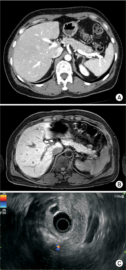
Fig. 1 Representative features of noninvasive radiologic findings of focal nesidioblastosis. Pancreatobiliary-protocol CT scanning (A), MRI (B), and endoscopic ultrasonography (C) showed no abnormal lesions in the pancreas of our patient.
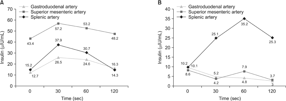
Fig. 2 Time-dependent changes of insulin concentraion in gastroduodenal artery, superior mesenteric artery and splenic artery, using selective intra-arterial calcium stimulation with hepatic vein sampling. (A) Our patient with focal nesidioblastosis had high insulin concentration in the superior mesenteric artery at our hospital. (B) At the previous hospital, insulin concentration was higher in the splenic artery than in the superior mesenteric artery and gastroduodenal artery.
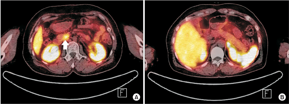
Fig. 3 Representative features of 68gallium- DOTA-D-Phe1-Tyr3-octreotide (DOTATOC) PET scanning of focal nesidioblastosis. (A) Increased DOTATOC uptake in the pancreas head area was found clearly (white arrow). (B) No other abnormal lesion was found in the rest of the pancreas.
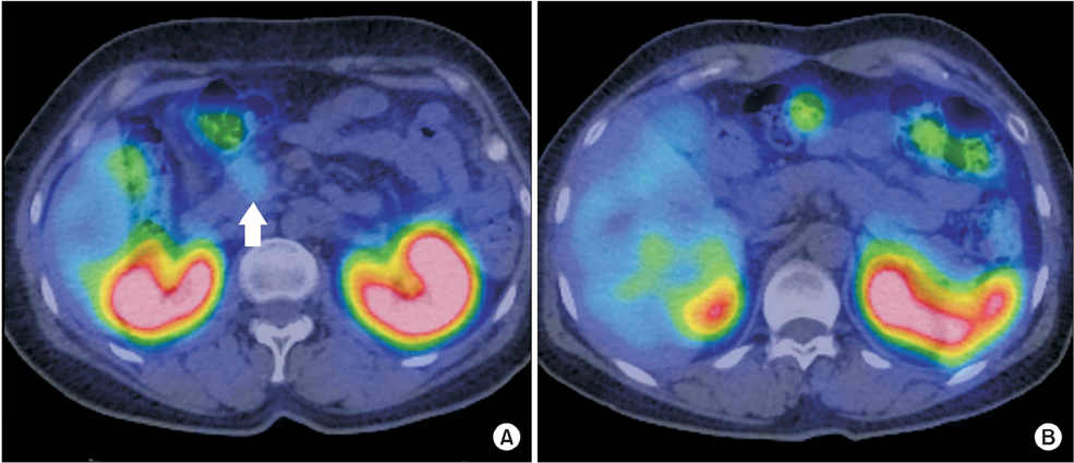
Fig. 4 Representative features of 111indium-pentetreotide diethylene triamine pentaacetic acid octreotide scanning of focal nesidioblastosis. (A) Increased octreotide uptake was found in the head of the pancreas, which was less clear than that of 68gallium- DOTA-D-Phe1-Tyr3-octreotide PET scanning (white arrow). (B) No other abnormal lesion was found in the rest of the pancreas.
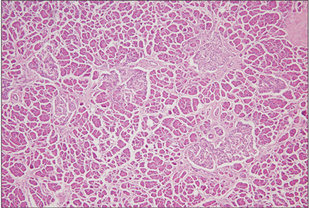
Fig. 5 Pathologic feature of focal hyperplasia of islets of Langerhans. Hyperplasia of ductulo-insular complex, nuclear hyperchromasia and β-cells enlargement was found with no localized aggregation (H&E, ×100).
Reference
-
1. Stanley CA, Lieu YK, Hsu BY, Burlina AB, Greenberg CR, Hopwood NJ, et al. Hyperinsulinism and hyperammonemia in infants with regulatory mutations of the glutamate dehydrogenase gene. N Engl J Med. 1998; 338:1352–1357.2. Laidlaw GF. Nesidioblastoma, the islet tumor of the pancreas. Am J Pathol. 1938; 14:125–134.3. Starke A, Saddig C, Kirch B, Tschahargane C, Goretzki P. Islet hyperplasia in adults: challenge to preoperatively diagnose noninsulinoma pancreatogenic hypoglycemia syndrome. World J Surg. 2006; 30:670–679.4. McElroy MK, Lowy AM, Weidner N. Case report: focal nesidioblastosis ("nesidioblastoma") in an adult. Hum Pathol. 2010; 41:447–451.5. Doppman JL, Shawker TH, Miller DL. Localization of islet cell tumors. Gastroenterol Clin North Am. 1989; 18:793–804.6. Guettier JM, Kam A, Chang R, Skarulis MC, Cochran C, Alexander HR, et al. Localization of insulinomas to regions of the pancreas by intraarterial calcium stimulation: the NIH experience. J Clin Endocrinol Metab. 2009; 94:1074–1080.7. Doppman JL, Miller DL, Chang R, Shawker TH, Gorden P, Norton JA. Insulinomas: localization with selective intraarterial injection of calcium. Radiology. 1991; 178:237–241.8. Srirajaskanthan R, Kayani I, Quigley AM, Soh J, Caplin ME, Bomanji J. The role of 68Ga-DOTATATE PET in patients with neuroendocrine tumors and negative or equivocal findings on 111In-DTPA-octreotide scintigraphy. J Nucl Med. 2010; 51:875–882.9. Buchmann I, Henze M, Engelbrecht S, Eisenhut M, Runz A, Schafer M, et al. Comparison of 68Ga-DOTATOC PET and 111In-DTPAOC (Octreoscan) SPECT in patients with neuroendocrine tumours. Eur J Nucl Med Mol Imaging. 2007; 34:1617–1626.10. Reubi JC, Schar JC, Waser B, Wenger S, Heppeler A, Schmitt JS, et al. Affinity profiles for human somatostatin receptor subtypes SST1-SST5 of somatostatin radiotracers selected for scintigraphic and radiotherapeutic use. Eur J Nucl Med. 2000; 27:273–282.
- Full Text Links
-
- Actions
-
Cited
- CITED
-
- Close
- Share
-
- Similar articles
-
- Ga68-DOTA Peptide PET/CT to Detect Occult Mesenchymal Tumor-Inducing Osteomalacia: A Case Series of Three Patients
- Radiolabeling of NOTA and DOTA with Positron Emitting 68Ga and Investigation of In Vitro Properties
- Chronologic Change of Hyperinsulinemic Hypoglycemic Disease-insulinoma and Nesidioblastosis in 64 Patients
- A Case of Focal Type Nesidioblastosis in Adult Treated with Distal Pancreatectomy
- Role of 68Ga-DOTATATE PET/CT in a Case of SDHB-Related Pterygopalatine Fossa Paraganglioma Successfully Controlled with Octreotide
