Malignant Oncocytoma of the Orbit: A Case Report
- Affiliations
-
- 1Department of Radiology, Chungnam National University Hospital, Chungnam National University School of Medicine, Daejeon, Korea. leeinho1974@hanmail.net
- 2Department of Ophthalmology, Chungnam National University Hospital, Chungnam National University School of Medicine, Daejeon, Korea.
- 3Department of Pathology, Chungnam National University Hospital, Chungnam National University School of Medicine, Daejeon, Korea.
Abstract
- Malignant oncocytoma of the orbit is extremely rare. A 76-year-old man presented with a 10-year history of a mass in the left orbit. Facial computed tomography revealed a large homogeneously enhancing mass. Magnetic resonance images showed a mass with isosignal intensity on a T1-weighted image and low signal intensity on a T2-weighted image. No restricted diffusion was observed on diffusion weighted imaging and apparent diffusion coefficient mapping. The patient underwent an incisional biopsy, and histopathological review revealed a malignant oncocytoma that likely originated in the caruncle of the left eye.
MeSH Terms
Figure
-
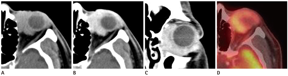
Fig. 1 76-year-old man with malignant oncocytoma of the left orbit. A. Non-contrast axial CT scan shows a relatively well-circumscribed, high-attenuated mass (77 HU) in the left orbit. B, C. Contrast-enhanced axial (B) and coronal (C) CT scans demonstrate strong enhancement (140 HU) in the left orbital mass. D. PET-CT scan shows high FDG uptake in the left orbital mass. Note.-FDG = fluorodeoxyglucose, HU = Hounsfield unit, PET = positron emission tomography

Fig. 2 The mass shows isosignal intensity on T1WI (A) and low signal intensity on T2WI (B). Note.-WI = weighted image

Fig. 3 Histopathologic examination (A and B; hematoxylin and eosin stain, × 100 and × 200) revealed the proliferation of atypical oncocytic neoplastic cells with stromal invasion, which is suggestive of malignant oncocytoma.
Reference
-
1. Kim SY, Paik JS, Yang SW. A case of oncocytoma of the caruncle. J Korean Ophthalmol Soc. 2010; 51:598–600.2. Radnót M. Aus Onkocyten bestehende adenomartige Hyperplasie in der Tränensackwand. Ophthalmologica. 1941; 101:95–100.3. Marglani O, Alherabi A, Corsten M. Malignant oncocytoma of the lacrimal sac with cervical metastasis: case report and literature review. J Otolaryngol Head Neck Surg. 2008; 37:E8–E10.4. Stefanyszyn MA, Hidayat AA, Pe'er JJ, Flanagan JC. Lacrimal sac tumors. Ophthal Plast Reconstr Surg. 1994; 10:169–184.5. Luthra CL, Doxanas MT, Green WR. Lesions of the caruncle: a clinicohistopathologic study. Surv Ophthalmol. 1978; 23:183–195.6. Biggs SL, Font RL. Oncocytic lesions of the caruncle and other ocular adnexa. Arch Ophthalmol. 1977; 95:474–478.7. Gonnering RS, Sonneland PR. Oncocytic carcinoma of the plica semilunaris with orbital extension. Ophthalmic Surg. 1987; 18:604–607.8. Yuen HK, Cheuk W, Cheng AC, Anh C, Chan N. Malignant oncocytoma of the lacrimal sac as an unusual cause of epiphora. Ophthal Plast Reconstr Surg. 2007; 23:70–72.9. Tan TJ, Tan TY. CT features of parotid gland oncocytomas: a study of 10 cases and literature review. AJNR Am J Neuroradiol. 2010; 31:1413–1417.10. Patel ND, van Zante A, Eisele DW, Harnsberger HR, Glastonbury CM. Oncocytoma: the vanishing parotid mass. AJNR Am J Neuroradiol. 2011; 32:1703–1706.
- Full Text Links
-
- Actions
-
Cited
- CITED
-
- Close
- Share
-
- Similar articles
-
- Renal Oncocytoma Presenting as a Huge Hypervascular Mass on Ultrasound
- A Case of Adrenocortical Oncocytoma Misrecognized for Fibrolamellar Hepatocellular Carcinoma
- A Case of Primary Malignant Lymphoma of the Orbit Treated by Radiotherapy
- A Case of Renal Oncocytoma with Synchronous Contralateral Renal Cell Carcinoma
- A case of renal oncocytoma
