Effects of implant thread profile on insertion stress generation in cortical bone studied by dynamic finite element simulation
- Affiliations
-
- 1Department of Orthodontics, School of Dentistry, Kyungpook National University, Daegu, Korea.
- 2Department of Prosthodontics, School of Dentistry, Kyungpook National University, Daegu, Korea. prosth95@knu.ac.kr
- KMID: 2321789
- DOI: http://doi.org/10.4047/jkap.2014.52.4.279
Abstract
- PURPOSE
The aim of this study was to investigate the effect of implant thread profile on the marginal bone stresses which develop during implant insertion.
MATERIALS AND METHODS
Four experimental implants were created by placing four different thread systems on the body (4.1 mm x 10 mm) of the ITI standard implant. The thread types studied in this study included the buttress, v-shape, reverse buttress, and square shape threads. In order to examine the insertion stress generation, 3D dynamic finite element analysis was performed which simulated the insertion process of implants into a 1.2 mm thick cortical bone plate (containing 3.5 mm pilot hole) using a PC-based DEFORM 3D (ver 6.1, SFTC, Columbus, OH, USA) program.
RESULTS
Insertion stresses higher than human cortical bone developed around the implants. The level of insertion stresses was much different depending on the thread. Stress level was lowest near the v-shape thread, and highest near the square shaped thread. Difference in the interfacial bone stress level was more noticeable near the valley than the tip of the threads.
CONCLUSION
Among the four threads, the v-shape thread was turned out to minimize the insertion stress level and thereby create better conditions for implant osseointegration.
Figure
-
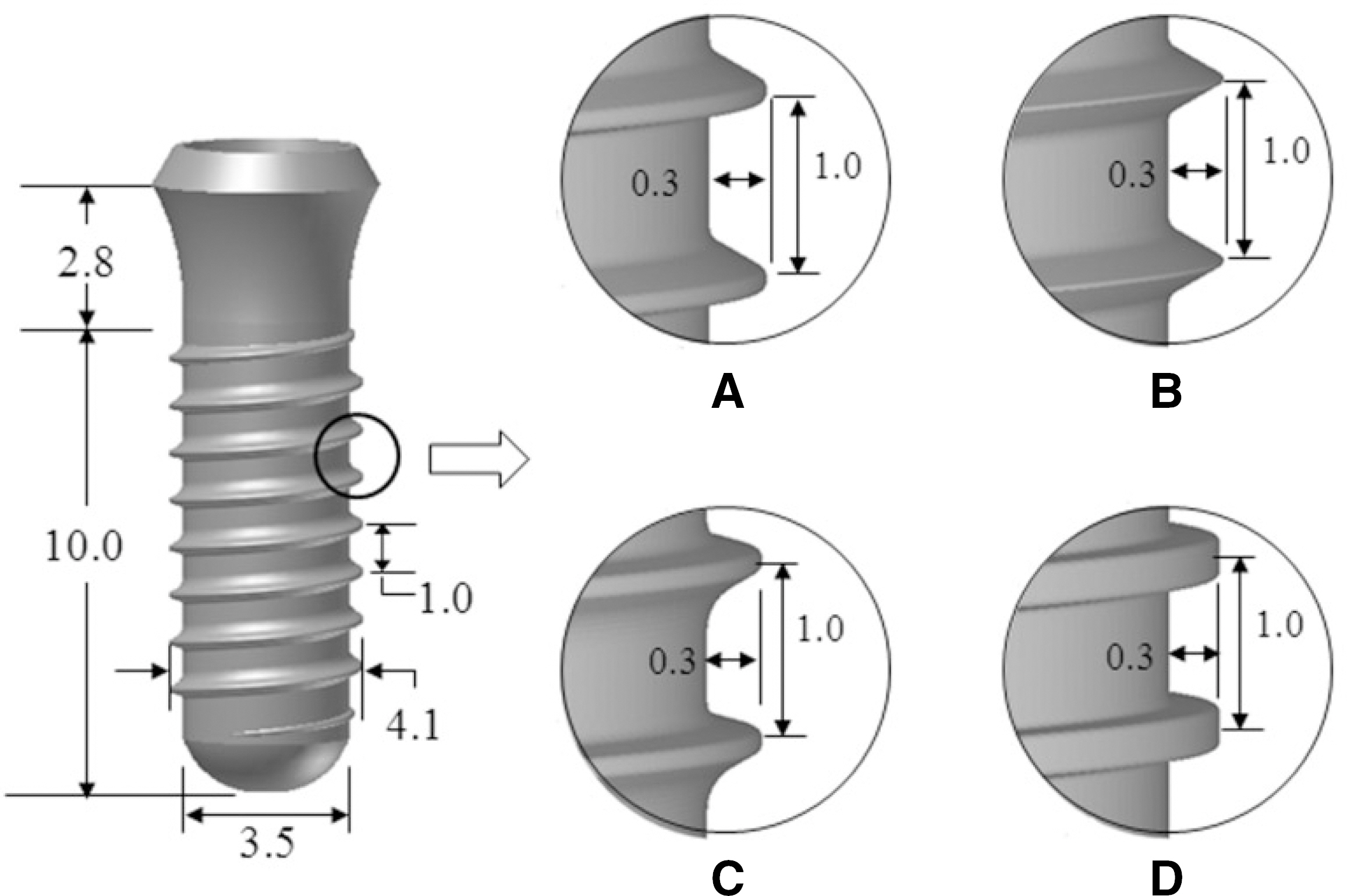
Fig. 1. Cylinder shaped implant body that incorporates four different thread profiles: (A) buttress thread, (B) V-shaped thread, (C) reverse buttress thread, and (D) square shape thread (unit: mm).

Fig. 2. Finite element method (FEM) simulation/analysis model consisting of implant (which was treated as a rigid material, and thus not meshed) and cortical bone mesh, shown with the axis system (unit: mm).
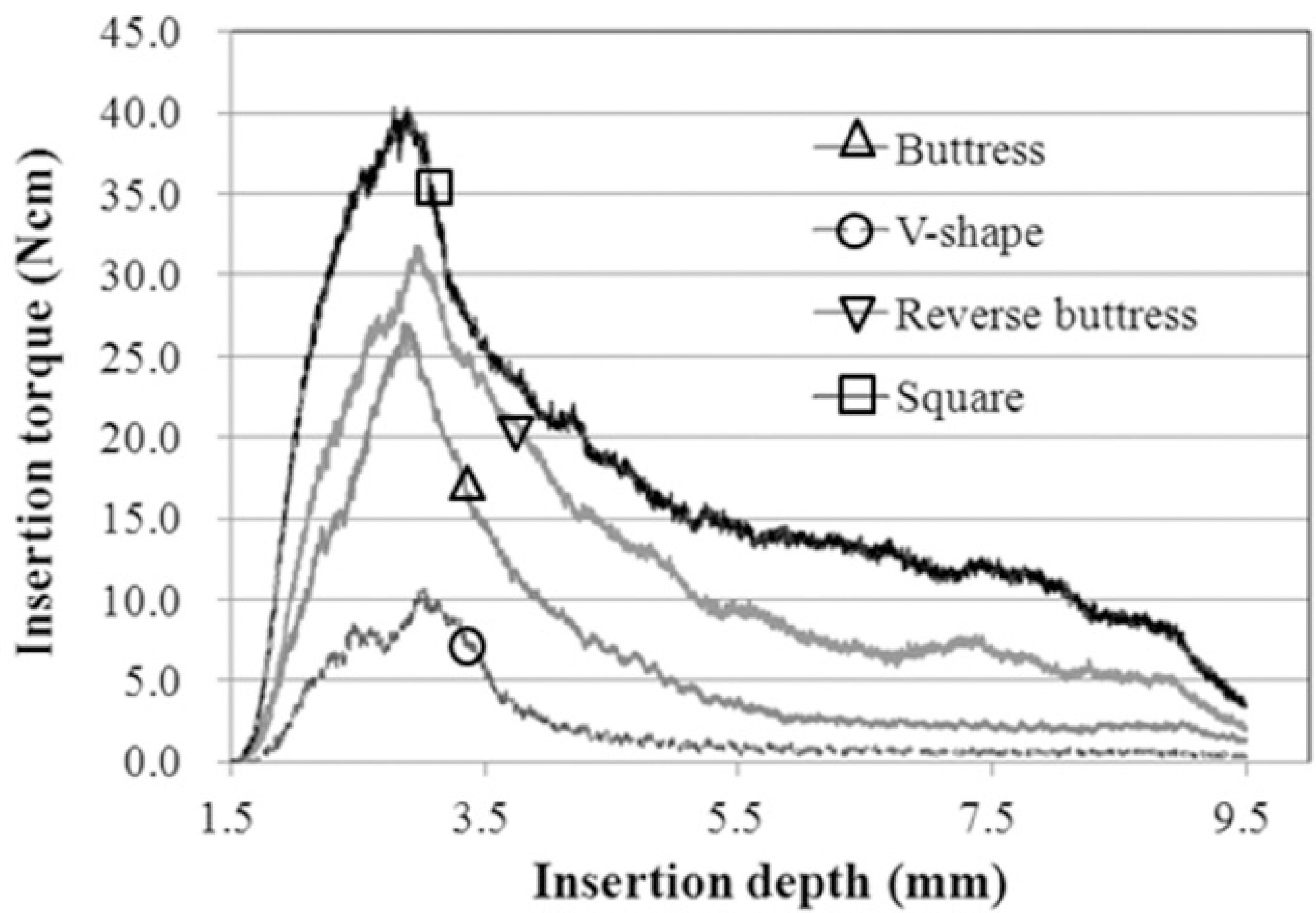
Fig. 3. The FEM calculated insertion torque when the implant incorporating each of 4 different threads is inserted into the 1.2 mm thick cortical bone.
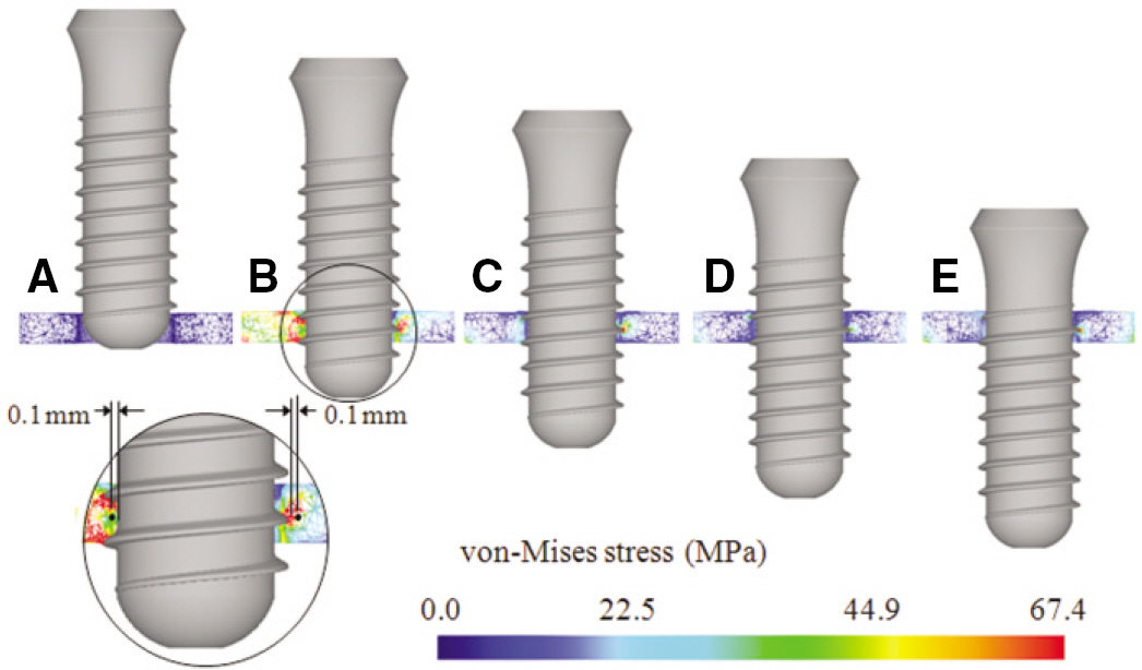
Fig. 4. Stress (von-Mises stress) development in the cortical bone around the implant which incorporates buttress threads; at (A) the initial stage (insertion depth d = 1.50 mm), (B) d = 3.5 mm, (C) d = 5.5 mm, (D) d = 7.5 mm, and (E) d = 9.5 mm. Two stress monitoring points are shown in (B) and (F) shows the stress band. Cut-off stress: 67.36 MPa (16.84 GPa ×4,000 μ -strain).

Fig. 5. Stress history recorded at the two stress monitoring points (shown in Fig. 4B), located 0.1 mm apart from (A) the thread tip, and (B) the thread valley.
Cited by 1 articles
-
Stress dissipation characteristics of four implant thread designs evaluated by 3D finite element modeling
Ok-Hyun Nam, Won-Jae Yu, Hee-Moon Kyung
J Korean Acad Prosthodont. 2015;53(2):120-127. doi: 10.4047/jkap.2015.53.2.120.
Reference
-
1. Cochran DL. A comparison of endosseous dental implant surfaces. J Periodontol. 1999; 70:1523–39.
Article2. Buser D, Schenk RK, Steinemann S, Fiorellini JP, Fox CH, Stich H. Influence of surface characteristics on bone integration of titanium implants. A histomorphometric study in miniature pigs. J Biomed Mater Res. 1991; 25:889–902.
Article3. Grunder U, Polizzi G, Goene′ R, Hatano N, Henry P, Jackson WJ, Kawamura K, Ko¨hler S, Renouard F, Rosenberg R, Triplett G, Werbitt M, Lithner B. A 3-year prospective multicenter follow-up report on the immediate and delayed-immediate placement of implants. Int J Oral Maxillofac Implants. 1999; 14:210–6.4. Berglundh T, Persson L, Klinge B. A systematic review of the incidence of biological and technical complications in implant dentistry reported in prospective longitudinal studies of at least 5 years. J Clin Periodontol. 2002; 29:197–212.5. Esposito M, Thomsen P, Ericson LE, Lekholm U. Histopathologic observations on early oral implant failures. Int J Oral Maxillofac Implants. 1999; 14:798–810.6. Friberg B, Jemt T, Lekholm U. Early failures in 4, 641 consec-utively placed Bra�nemark dental implants: a study from stage 1 surgery to the connection of completed prostheses. Int J Oral Maxillofac Implants. 1991; 6:142–6.7. Sakka S, Baroudi K, Nassani MZ. Factors associated with early and late failure of dental implants. J Investig Clin Dent. 2012; 3:258–61.
Article8. Pye AD, Lockhart DE, Dawson MP, Murray CA, Smith AJ. A review of dental implants and infection. J Hosp Infect. 2009; 72:104–10.9. Miyamoto Y. Risk factors of dental implant failure and the counter meausres. To decrease the implant failures. Shikoku Dent Res. 2006; 18:211–20.10. Manor Y, Oubaid S, Mardinger O, Chaushu G, Nissan J. Characteristics of early versus late implant failure: a retrospective study. J Oral Maxillofac Surg. 2009; 67:2649–52.
Article11. Pjetursson BE, Tan K, Lang NP, Bra¨gger U, Egger M, Zwahlen M. A systematic review of the survival and complication rates of fixed partial dentures (FPDs) after an observation period of at least 5 years. Clin Oral Implants Res. 2004; 15:625–42.12. Jung RE, Pjetursson BE, Glauser R, Zembic A, Zwahlen M, Lang NP. A systematic review of the 5-year survival and complication rates of implant-supported single crowns. Clin Oral Implants Res. 2008; 19:119–30.
Article13. Lang NP, Mombelli A, Tonetti MS, Bra¨gger U, Ha¨mmerle CH. Clinical trials on therapies for peri-implant infections. Ann Periodontol. 1997; 2:343–56.
Article14. Piattelli A, Scarano A, Piattelli M. Histologic observations on 230 retrieved dental implants: 8 years' experience (1989-1996). J Periodontol. 1998; 69:178–84.
Article15. Tada S, Stegaroiu R, Kitamura E, Miyakawa O, Kusakari H. Influence of implant design and bone quality on stress/strain distribution in bone around implants: a 3-dimensional finite element analysis. Int J Oral Maxillofac Implants. 2003; 18:357–68.16. Petrie CS, Williams JL. Comparative evaluation of implant designs: influence of diameter, length, and taper on strains in the alveolar crest. A three-dimensional finite-element analysis. Clin Oral Implants Res. 2005; 16:486–94.17. Ha¨nggi MP, Ha¨nggi DC, Schoolfield JD, Meyer J, Cochran DL, Hermann JS. Crestal bone changes around titanium implants. Part I: A retrospective radiographic evaluation in humans comparing two non-submerged implant designs with different machined col-lar lengths. J Periodontol. 2005; 76:791–802.18. Clelland NL, Gilat A. The effect of abutment angulation on stress transfer for an implant. J Prosthodont. 1992; 1:24–8.
Article19. Gotfredsen K, Berglundh T, Lindhe J. Bone reactions adjacent to titanium implants with different surface characteristics subjected to static load. A study in the dog (II). Clin Oral Implants Res. 2001; 12:196–201.
Article20. O'Brien GR, Gonshor A, Balfour A. A 6-year prospective study of 620 stress-diversion surface (SDS) dental implants. J Oral Implantol. 2004; 30:350–7.21. Chun HJ, Shin HS, Han CH, Lee SH. Influence of implant abutment type on stress distribution in bone under various loading conditions using finite element analysis. Int J Oral Maxillofac Implants. 2006; 21:195–202.22. Hansson S. The implant neck: smooth or provided with retention elements. A biomechanical approach. Clin Oral Implants Res. 1999; 10:394–405.
Article23. Misch CE. Dental implant prosthetics. St. Louis: Mosby;2005. p. 322–47.24. Chun HJ, Cheong SY, Han JH, Heo SJ, Chung JP, Rhyu IC, Choi YC, Baik HK, Ku Y, Kim MH. Evaluation of design parameters of osseointegrated dental implants using finite element analysis. J Oral Rehabil. 2002; 29:565–74.
Article25. Hansson S, Werke M. The implant thread as a retention element in cortical bone: the effect of thread size and thread profile: a finite element study. J Biomech. 2003; 36:1247–58.
Article26. Kong L, Hu K, Li D, Song Y, Yang J, Wu Z, Liu B. Evaluation of the cylinder implant thread height and width: a 3-dimensional finite element analysis. Int J Oral Maxillofac Implants. 2008; 23:65–74.27. Seo YH, Vang MS, Yang HS, Park SW, Park HO, Lim HP. Three dimentional finite element analysis of stress distribution for different implant thread slope. J Korean Acad Prosthodont. 2007; 45:482–91.28. Yu W, Park HS, Kyung HM, Kwon OW. Dynamic simulation of the self-tapping insertion process of orthodontic microimplants into cortical bone with a 3-dimensional finite element method. Am J Orthod Dentofacial Orthop. 2012; 142:834–41.
Article29. Frost HM. Wolff's Law and bone's structural adaptations to mechanical usage: an overview for clinicians. Angle Orthod. 1994; 64:175–88.30. Meyer U, Vollmer D, Runte C, Bourauel C, Joos U. Bone loading pattern around implants in average and atrophic edentulous maxillae: a finite-element analysis. J Maxillofac Surg. 2001; 29:100–5.
Article31. Isidor F. Loss of osseointegration caused by occlusal load of oral implants. A clinical and radiographic study in monkeys. Clin Oral Implants Res. 1996; 7:143–52.
Article
- Full Text Links
-
- Actions
-
Cited
- CITED
-
- Close
- Share
-
- Similar articles
-
- Influence of microthread design on marginal cortical bone strain developement: A finite element analysis
- Three-dimensional finite element analysis for stress distribution on the diameter of orthodontic mini-implants and insertion angle to the bone surface
- The three dimensional finite element analysis of the stress distribution according to the implant and thread designs
- Three-dimensional finite element analysis according to the insertion depth of an immediately loaded implant in the anterior maxilla
- Finite element analysis of cortical bone strain induced by self-drilling placement of orthodontic microimplant
