J Korean Acad Prosthodont.
2010 Apr;48(2):135-142.
Evaluation using Replica Technique on the marginal and internal fitness of zirconia cores by several CAD/CAM systems
- Affiliations
-
- 1Department of Advanced Prosthetic Dentistry, Graduate School of Clinical Dentistry, Korea University, Seoul, Korea. swshin@korea.ac.kr
Abstract
- PURPOSE
This study was aimed to compare the margin and internal fitness of single anterior all-ceramic crown zirconia core made by three deferent CAD/CAM systems. MATERIAL AND METHODS: Five single zirconia cores were manufactured by three deferent CAD/CAM systems(Cerasys(R)system, KaVo Everest(R)system, Lava(TM) system). The manufactured zirconia cores were duplicated through the use of replica technique, and a replicated sample was sectioned in the center of bucolingual and mesiodistal direction to measure the marginal and internal gap. Measurement was carried out by using measuring microscope (AXIO(R)) and I-Solution(R) and analysed through the use of ANOVA.
RESULTS
As for the mean marginal fitness of the zirconia core, it was 84.74 +/- 27.57 micrometer, in Cerasys(R), 80.23 +/- 21.07 micrometer in KaVo Everest(R) and 96.37 +/- 11.45 micrometer in Lava(TM), and as for the mean internal gap, it was 94.11 +/- 30.07 micrometer in Cerasys(R), 92.31 +/- 25.18 micrometer in KaVo Everest(R), and 94.99 +/- 18.74 micrometer in Lava(TM). There was no significant statistically deference among the total average gap of three systems. The internal gap in KaVo Everest(R) seemed to be smaller than Lava(TM) (P < .05). The internal gap in the incisal area was larger in all of the three systems.
CONCLUSION
There was no difference in marginal fitness in Cerasys(R), KaVo Everest(R) and Lava(TM). As for the internal fitness, it was smaller in KaVo Everest(R) system than Lava(TM)system. In all of the three systems, there was a larger gap in incisal area. The marginal and internal gap was within the clinically allowed range in all of the three systems.
Keyword
MeSH Terms
Figure
-
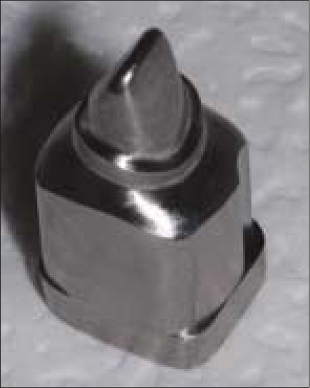
Fig. 1. Master models with Titanium Block made by using CAD/CAM system (Addtech Co., Seoul, Korea).
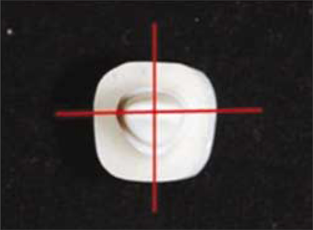
Fig. 2. Directions and positions of sectioning the replica materials.
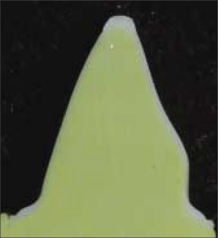
Fig. 3. Surface aspect after sectioning with buccolingual direction.
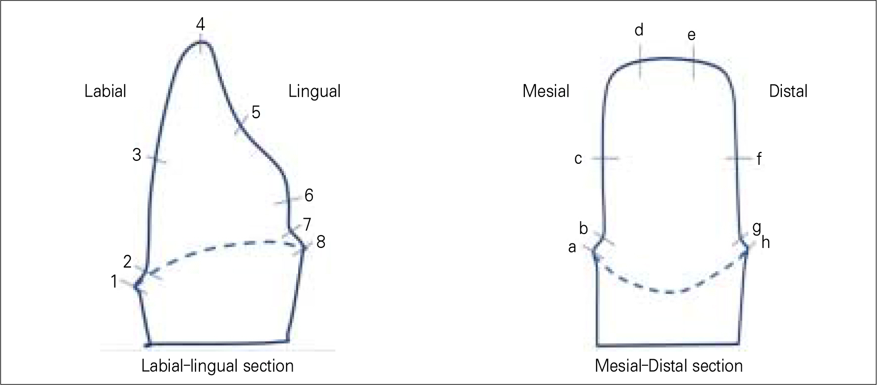
Fig. 4. Reference points to measure the thickness.
Reference
-
1.Andersson M., Razzoog ME., Ode ′n A., Hegenbarth EA., Lang BR. Procera: a new way to achieve an all-ceramic crown. Quintessence Int. 1998. 29:285–96.2.Boening KW., Wolf BH., Schmidt AE., Ka ¨stver K., Walter MH. Clinical fit of Procera Allceram crowns. J Prosthet Dent. 2000. 84:419–24.
Article3.Seghi PR., Sorensen JA. Relative flexural strength of six new ceramic materials. Int J Prosthodont. 1995. 8:239–46.4.Sturdevant JR., Bayne SC., Heymann HO. Margin gap size of ceramic inlays using second-generation CAD/CAM equipment. J Esthet Dent. 1999. 11:206–14.
Article5.Tinschert J., Natt G., Mautsch ., Spikermann H., Anusavice KJ. Marginal fit of alumina-and zirconia-based fixed partial dentures produced by a CAD/CAM system. Oper Dent. 2001. 26:367–74.6.Lawn BR., Deng Y., Thompson VP. Use of contact testing in the characterization and design of all-ceramic crown-like layer structures: a review. J Prosthet Dent. 2001. 86:495–510.
Article7.Jeon MH., Jeon YC., Jeong CM., Lim JS., Jeong HC. A study of precise fit of the CAM zirconia all-ceramic framework. J Korean Acad Prosthodont. 2005. 43:611–21.8.Seo JY., Park IN., Lee KW. Fracture strength between different connector designs of zirconia core for posterior fixed partial dentures manufactured with CAD/CAM system. J Korean Acad Prosthodont. 2006. 44:29–39.9.Bader J., Rozier R., McFall W Jr., Ramsey D. Effect of crown margins on periodontal conditions in regularly attending patients. J Prosthet Dent. 1991. 65:75–9.
Article10.Grasso J., Nalbandian J., Sanford C., Baili H. Effect of restoration quality on periodontal health. J Prosthet Dent. 1985. 53:14–9.
Article11.Schwartz N., Whitsett L., Berry T., Stewart J. Unserviceable crowns and fixed partial dentures: lifespan and causes for loss of serviceability. J Am Dent Assoc. 1970. 81:1395–401.
Article12.Felton D., Kanoy B., Bayne S., Wirthman G. Effect of in vivo crown margin discrepancies on periodontal health. J Prosthet Dent. 1991. 65:357–64.
Article13.Walton J., Gardner F., Agar J. A survey of crown and fixed partial denture failures: length of service and reasons for replacement. J Prosthet Dent. 1986. 56:416–21.
Article14.Jorgensen KD. Factors affecting the film thickness of zinc phosphate cement. Acta Odontol Scan. 1960. 18:479–90.15.Council on dental materials and devices. Revised american national standards institute/American dental association specification No. 8 for zinc phosphate cement. J Am Dent Assoc. 1978. 96:121–3.16.Christensen GJ. Marginal fit of gold castings. J Prosthet Dent. 1966. 16:297–305.17.McLean JW., von Fraunhofer JA. The estimation of cement film thickness by an in vivo technique. Br Dent J. 1971. 131:107–11.18.McLean JW., von Fraunhofer JA. Polycarboxylate cements.: Five years' experience in general practice. Br Dent J. 1972. 132:9–15.
Article19.Hung SH., Hung KS., Eick JD., Chappel RP. Marginal fit of porcelain-fused-to metal and two types of ceramic crown. J Prosthet Dent. 1990. 63:26–31.20.May KB., Russell MM., Razzoog ME., Lang BR. Precision of fit: the procera Allceram crown. J Prosthet Dent. 1998. 80:394–404.
Article21.Hertlein G., Hoscheler S., Frank S., Suttor D. Marginal fit of CAD/CAM manufactured all ceramic prosthesis. J Dent Res. 2001. 80:42–4.22.Kim DK., Cho IH., Lim JH., Lim HS. On the marginal fidelity of all-ceramic core using CAD/CAM system. J Korean Acad Prosthodont. 2003. 41:20–34.23.Yang JH., Yeo IS., Lee SH., Han JS., Lee JB. Marginal fit of Celay/In-Ceram, conventional In-Ceram and Empress 2 all-ceramic single crowns. J Korean Acad Prosthodont. 2002. 40:131–9.24.Carter JM., Sorensen SE., Johnson RR., Teitelbaum RL., Levine MS. Punch shear testing of extracted vital and endodontically treated teeth. J Biomech. 1983. 16:841–8.
Article25.Strawn SE., White JM., Marshall GW., Gee L., Goodis HE., Marchall SJ. Spectroscopic changes in human dentin exposed to various storage solution-short term. J Dent. 1996. 24:417–23.26.Pera P., Bassi F., Carossa S. In vitro marginal adaptation of alumina porcelain ceramic crown. J Prosthet Dent. 1994. 72:585–90.27.Koo JY., Lim JH., Cho IH. Marginal fidelity according to the margin types of all ceramic crowns. J Korean Acad Prosthodont. 1997. 35:445–57.28.Sorensen JA. A standardized method for determination of crown margin. J Prosthet Dent. 1990. 64:18–24.29.Moon BH., Yang JH Lee SH., Chung HY. A study on the marginal fit of all-ceramic crown using ccd camera. J Korean Acad Prosthodont. 1998. 36:273–92.30.Molin M., Karlsson S. The fit of gold inlays and three ceramic inlay system. Aclinical and in vitro study. Acta Odontol Scand. 1993. 51:201–16.31.Habib Y., Georges E., Salim M., Albert S., Khaldoun T. In vitro evaluation of the “Replica Technique” in the measurement of the fit of Procera crown. J Contemp Dent Pract. 2008. 9:25–32.32.Davis DR. Comparison of fit of two types of all ceramic crowns. J Prosthet Dent. 1988. 59:13–6.33.Abbate MF., Tjan A., Fox WM. Comparison of marginal fit of various ceramic crown systems. J Prosthet Dent. 1989. 61:527–31.34.Belser UC., Mecentee MI., Richter WA. Fit of three porcelain-fusedto metal marginal designs in vivo: a scanning electron microscope study. J Prosthet Dent. 1985. 53:24–9.35.Bindle A., Mormann WH. Marginal and internal fit of allceramic CAD/CAM crown-coping on chamfer preparations. J Oral Rehabil. 2005. 32:441–7.36.Valderrama S., Roekel NV., Andersson M., Goodacre CJ., Munoz CA. A comparison of the marginal and internal adaptation of titanium and gold-platinum-palladium metal ceramic crowns. Int J Prosthodont. 1995. 8:29–37.
- Full Text Links
-
- Actions
-
Cited
- CITED
-
- Close
- Share
-
- Similar articles
-
- Marginal and internal fitness of three-unit zirconia cores fabricated using several CAD/CAM systems
- Comparative study in marginal adaptation of zirconia cores fabricated with 3 different CAD/CAM systems
- The fit of zirconia core fabricated with CAD/CAM system
- Comparison of the fit accuracy of zirconia-based prostheses generated by two CAD/CAM systems
- Marginal and internal fit of all ceramic crown using the replica technique and the triple-scan protocol
