A Case of Pulmonary Artery Sarcoma Presented as Cavitary Pulmonary Lesions
- Affiliations
-
- 1Department of Internal Medicine, Bundang CHA Medical Center, CHA University College of Medicine, Seongnam, Korea. imekkim@cha.ac.kr
Abstract
- Pulmonary artery sarcoma (PAS) is a rare, poorly differentiated malignancy arising from the intimal layer of the pulmonary artery. Contrast-enhanced chest computed tomography (CT) is a good diagnostic modality that shows a low-attenuation filling defect of the pulmonary artery in PAS patients. An 18-year-old man was referred to our hospital for the evaluation and management of cavitary pulmonary lesions that did not respond to treatment. A contrast-enhanced CT of the chest was performed, which showed a filling defect within the right interlobar pulmonary artery. The patient underwent a curative right pneumonectomy after confirmation of PAS. Although lung parenchymal lesions of PAS are generally nonspecific, it can be presented as cavities indicate pulmonary infarcts. Clinicians must consider the possibility of PAS as well as pulmonary thromboembolism in patients with pulmonary infarcts. So, we report the case with PAS that was diagnosed during the evaluation of cavitary pulmonary lesions and reviewed the literatures.
Keyword
MeSH Terms
Figure
-
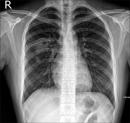
Figure 1 Initial chest radiography shows patchy consolidations in the right upper lobe.
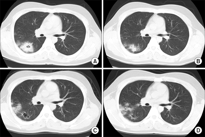
Figure 2 Non-enhanced chest computed tomography (CT) images. (A) Initial non-enhanced chest CT shows a 5-cm cavitary consolidation in the right upper lobe posterior segment. (B-D) Follow-up CT show changes in the pulmonary lesions, decrease in size of the main cavitary consolidation and increase in the number and size of adjacent nodular consolidations.
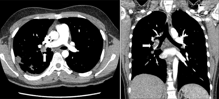
Figure 3 Contrast-enhanced chest computed tomography shows a filling defect within the lumen of the right interlobar pulmonary artery (arrows).
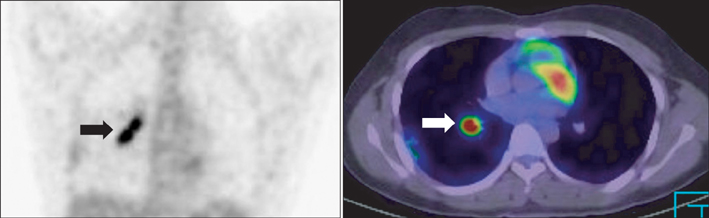
Figure 4 Positron emission tomography-computed tomography shows increased 18F-fluorodeoxyglucose uptake, with a maximum standardized uptake value of 9.6 along the right interlobar pulmonary artery (arrows).
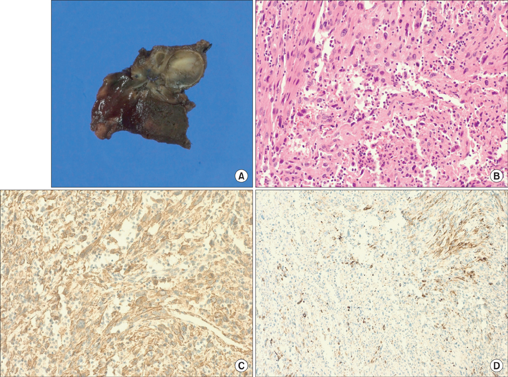
Figure 5 (A) Intravascular mass. (B-D) Histopathologic examination reveals abundant spindle cells with high cellularity, frequent mitoses, and nuclear pleomorphism (B, H&E stain, ×20). Immunohistochemical staining is positive for smooth-muscle actin (C, ×20) and desmin (D, ×10).
Reference
-
1. Mandelstamm M. Über primäre Neubildungen des Herzens. Virchows Arch Pathol Anat Physiol Klin Med. 1923; 245:43–54.2. Blackmon SH, Rice DC, Correa AM, Mehran R, Putnam JB, Smythe WR, et al. Management of primary pulmonary artery sarcomas. Ann Thorac Surg. 2009; 87:977–984.3. Mussot S, Ghigna MR, Mercier O, Fabre D, Fadel E, Le Cesne A, et al. Retrospective institutional study of 31 patients treated for pulmonary artery sarcoma. Eur J Cardiothorac Surg. 2013; 43:787–793.4. Fukuda W, Morohashi S, Fukuda I. Intimal sarcoma of the pulmonary artery: diagnostic challenge. Acta Cardiol. 2011; 66:539–541.5. Farooki ZQ, Chang CH, Jackson WL, Clapp SK, Hakimi M, Arciniegas E, et al. Primary pulmonary artery sarcoma in two children. Pediatr Cardiol. 1988; 9:243–251.6. Gadkowski LB, Stout JE. Cavitary pulmonary disease. Clin Microbiol Rev. 2008; 21:305–333.7. Kim NR, Han J. Pathologic review of cystic and cavitary lung diseases. Korean J Pathol. 2012; 46:407–414.8. Delany SG, Doyle TC, Bunton RW, Hung NA, Joblin LU, Taylor DR. Pulmonary artery sarcoma mimicking pulmonary embolism. Chest. 1993; 103:1631–1633.9. Mattoo A, Fedullo PF, Kapelanski D, Ilowite JS. Pulmonary artery sarcoma: a case report of surgical cure and 5-year follow-up. Chest. 2002; 122:745–747.10. Ishiguro T, Kasahara K, Matsumoto I, Waseda R, Minato H, Kimura H, et al. Primary pulmonary artery sarcoma detected with a pulmonary infarction. Intern Med. 2007; 46:601–604.11. Nagai T, Tabata H, Uehata A. Primary sarcoma of pulmonary artery resembling large pulmonary thrombus: diagnostic utility of different imaging modalities. Eur Heart J. 2012; 33:2271.12. Ito K, Kubota K, Morooka M, Shida Y, Hasuo K, Endo H, et al. Diagnostic usefulness of 18F-FDG PET/CT in the differentiation of pulmonary artery sarcoma and pulmonary embolism. Ann Nucl Med. 2009; 23:671–676.13. Anderson MB, Kriett JM, Kapelanski DP, Tarazi R, Jamieson SW. Primary pulmonary artery sarcoma: a report of six cases. Ann Thorac Surg. 1995; 59:1487–1490.14. Hoiczyk M, Iliodromitis K, Bauer S, Konorza T, Philipp S, Bankfalvi A, et al. Intimal sarcoma of the pulmonary artery with unusual findings: a case report. Clin Res Cardiol. 2012; 101:397–401.15. Jamieson SW. Pulmonary artery sarcoma. Eur J Cardiothorac Surg. 2013; 43:793–794.
- Full Text Links
-
- Actions
-
Cited
- CITED
-
- Close
- Share
-
- Similar articles
-
- Surgical Treatment of Pulmonary Artery Sarcoma: One case report
- Pulmonary Artery Intimal Sarcoma with Lung Metastasis
- Pulmonary Artery Intimal Sarcoma Involving the Peripheral Pulmonary Artery, Initially Misdiagnosed as Pulmonary Artery Thromboembolism and Vasculitis: A Case Report
- Differential Imaging Features of Pulmonary Artery Dissection from Other Intraluminal Diseases of Pulmonary Artery: Two Cases Report
- A Case of Primary Pulmonary Artery Sarcoma Mimicking Pulmonary Embolism: Role of PET/CT for Differential Diagnosis
