Tuberc Respir Dis.
2014 Jan;76(1):34-37.
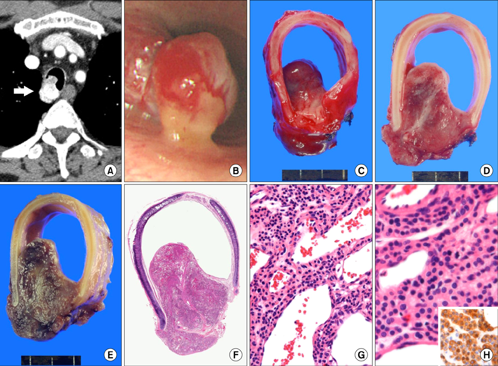
Two Cases of Glomus Tumor Arising in Large Airway: Well Organized Radiologic, Macroscopic and Microscopic Findings
- Affiliations
-
- 1Department of Pathology, Samsung Medical Center, Sungkyunkwan University School of Medicine, Seoul, Korea. hanjho@skku.edu
- 2Department of Thoracic Surgery, Samsung Medical Center, Sungkyunkwan University School of Medicine, Seoul, Korea.
Abstract
- Glomus tumors of the lung are rare benign neoplasm, originating from modified smooth muscle cells. The patients are usually presented with no or non-specific symptoms such as cough, dyspnea or hemoptysis. Although surgical treatment is considered as the treatment of choice, the endobronchial therapy can be applied to the patients who are unfit for surgical excision. Herein, we describe two rare cases of glomus tumor originated at large airway (trachea and main bronchus) without respiratory symptoms and review their characteristic radiologic, macroscopic and pathological features.
Keyword
Figure
Reference
-
1. Shugart RR, Soule EH, Johnson EW Jr. Glomus Tumor. Surg Gynecol Obstet. 1963; 117:334–340.2. Miettinen M, Paal E, Lasota J, Sobin LH. Gastrointestinal glomus tumors: a clinicopathologic, immunohistochemical, and molecular genetic study of 32 cases. Am J Surg Pathol. 2002; 26:301–311.3. Albores-Saavedra J, Gilcrease M. Glomus tumor of the uterine cervix. Int J Gynecol Pathol. 1999; 18:69–72.4. Gokten N, Peterdy G, Philpott T, Maluf HM. Glomus tumor of the ovary: report of a case with immunohistochemical and ultrastructural observations. Int J Gynecol Pathol. 2001; 20:390–394.5. Kim MJ, Sung WJ. Primary pulmonary glomus tumor, diagnosed by preoperative needle biopsy: report of one case and literature review. Korean J Pathol. 2008; 42:37–40.6. Ariizumi Y, Koizumi H, Hoshikawa M, Shinmyo T, Ando K, Mochizuki A, et al. A primary pulmonary glomus tumor: a case report and review of the literature. Case Rep Pathol. 2012; 2012:782304.7. Lee EW, Kim SO, Oh IJ, Ju JY, Cho GJ, Kim KS, et al. A case of bronchial glomus tumor. Tuberc Respir Dis. 2002; 53:445–449.8. Jeffery PK. Remodeling in asthma and chronic obstructive lung disease. Am J Respir Crit Care Med. 2001; 164(10 Pt 2):S28–S38.9. Ko JM, Jung JI, Park SH, Lee KY, Chung MH, Ahn MI, et al. Benign tumors of the tracheobronchial tree: CT-pathologic correlation. AJR Am J Roentgenol. 2006; 186:1304–1313.10. Grillo HC, Mathisen DJ. Primary tracheal tumors: treatment and results. Ann Thorac Surg. 1990; 49:69–77.11. Akata S, Yoshimura M, Park J, Okada S, Maehara S, Usuda J, et al. Glomus tumor of the left main bronchus. Lung Cancer. 2008; 60:132–135.12. Glazebrook KN, Laundre BJ, Schiefer TK, Inwards CY. Imaging features of glomus tumors. Skeletal Radiol. 2011; 40:855–862.


