Tuberc Respir Dis.
2009 Nov;67(5):467-470.
A Case of Post-Traumatic Pulmonary Pseudocyst Mimicking Pulmonary Cavitary Tuberculosis
- Affiliations
-
- 1Department of Internal Medicine, The Catholic University of Korea, College of Medicine, Seoul, Korea. mdlee@catholic.ac.kr
- 2Department of Radiology, The Catholic University of Korea, College of Medicine, Seoul, Korea.
Abstract
- A traumatic pulmonary pseudocyst is a rare complication of blunt thoracic trauma. The clinical symptoms and signs are similar to other respiratory diseases, such as pulmonary tuberculosis. Therefore, a trauma history with the resulting radiologic and clinical findings is important for making a diagnosis. A 26-year-old male was admitted to our hospital due to cough for 3 days. The chest x-ray revealed diffuse infiltrations and a cavitary lesion at the left lung. His left chest had hit a tree as a result of motorcycle accident one day before admission. Initially, it was assumed that his symptoms and chest X-ray might be due to a tuberculosis infection. However, bronchoscopy revealed old blood clots at both lungs, particularly in the left lower lobe bronchus. A transbronchial lung biopsy showed alveolar hemorrhage. A traumatic pulmonary pseudocyst was diagnosed from his trauma history and these findings. Computed tomography of the chest performed 4 months later showed regression of the cavitary lesion.
Keyword
MeSH Terms
Figure
-
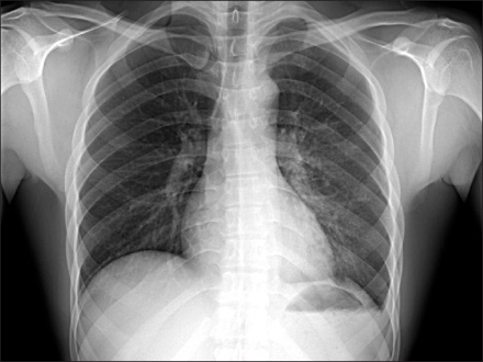
Figure 1 Initial posteroanterior chest X-ray shows ill-defined ground glass opacity and cavitary change in left lung field.
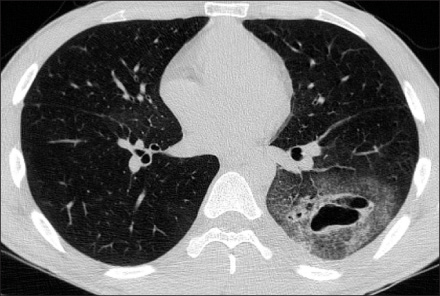
Figure 2 Computed tomography image with a lung window setting shows clustered small and large cavitary lesions with wall thickening in left lower lobe, predominantly superior segment.
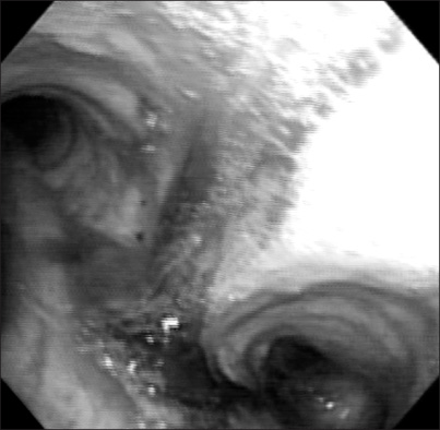
Figure 3 Flexible bronchoscopy shows old blood clot from trachea to both main bronchus, predominantly in left lower lobe bronchus.
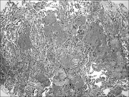
Figure 4 Microscopic finding shows intra-alveolar hemorrhage, deposit of fibrinoid fluid and focal infiltration of neutrophils (H&E stain, ×).
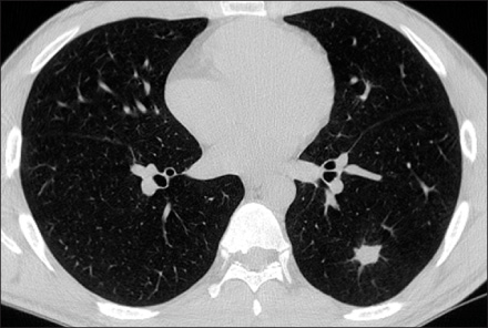
Figure 5 Follow up chest computed tomography performed 4 months later shows much regression of the previously noted cavitary and infiltrative lesion.
Reference
-
1. Kato R, Horinouchi H, Maenaka Y. Traumatic pulmonary pseudocyst. Report of twelve cases. J Thorac Cardiovasc Surg. 1989. 97:309–312.2. Ganske JG, Dennis DL, Vanderveer JB Jr. Traumatic lung cyst: case report and literature review. J Trauma. 1981. 21:493–496.3. Melloni G, Cremona G, Ciriaco P, Pansera M, Carretta A, Negri G, et al. Diagnosis and treatment of traumatic pulmonary pseudocysts. J Trauma. 2003. 54:737–743.4. Stulz P, Schmitt HE, Hasse J, Grädel E. Traumatic pulmonary pseudocysts and paramediastinal air cyst: two rare complications of blunt chest trauma. J Trauma. 1984. 24:850–853.5. Fagan CJ. Traumatic lung cyst. Am J Roentgenol Radium Ther Nucl Med. 1966. 97:186–194.6. Fagan CJ, Swischuk LE. Traumatic lung and paramediastinal pneumatoceles. Radiology. 1976. 120:11–18.7. Athanassiadi K, Gerazounis M, Kalantzi N, Kazakidis P, Fakou A, Kourousis D. Primary traumatic pulmonary pseudocysts: a rare entity. Eur J Cardiothorac Surg. 2003. 23:43–45.8. Sorsdahl OA, Powell JW. Cavitary pulmonary lesions following nonpenetrating chest trauma in children. Am J Roentgenol Radium Ther Nucl Med. 1965. 95:118–124.9. Lee SY, Lee SJ, Park HJ, Lee CS, Lee KR, Oh MH. A case of bilateral traumatic pulmonary pseudocyst. J Korean Soc Traumatol. 2004. 17:238–242.
- Full Text Links
-
- Actions
-
Cited
- CITED
-
- Close
- Share
-
- Similar articles
-
- A Case of Bronchial Artery Aneurysm in Cavitary Pulmonary Tuberculosis
- A Case of Chronic Necrotizing Pulmonary Aspergillosis Obscured by Cavitary Pulmonary Tuberculosis
- A Cavitary Lesion Changed to Pulmonary Nodule
- Clinical Courses of Cavitary Lesions in Pulmonary Tuberculosis
- Clinical Courses of Cavitary Lesions in Pulmonary Tuberculosis
