Tuberc Respir Dis.
2009 Jul;67(1):37-41.
A Case of Lymphocytic Interstitial Pneumonia Manifested as a Multi-focal Consolidation
- Affiliations
-
- 1Department of Internal Medicine, Hanyang University College of Medicine, Seoul, Korea. drterry@hanyang.ac.kr
- 2Department of Pathology, Hanyang University College of Medicine, Seoul, Korea.
Abstract
- Lymphocytic interstitial pneumonia (LIP) is a rare disorder characterized by a diffuse infiltration of the alveolar space, interstitium by lymphocytes, plasma cells, and reticuloendothelial cells. Although its etiology is unknown, LIP has been associated with autoimmune disorders and with viral infections. Because it's clinical and radiographic features are nonspecific, a confirmatory diagnosis is performed by open lung biopsy. A 59-year-old female presented dry cough, which had been present for 1 month. On initial findings of multifocal consolidation at the right middle lobe on both lower lobes in chest radiography, the first diagnosis of cryptogenic organizing pneumonia was suggested. On open lung biopsy, LIP was diagnosed. The patient had no autoimmune disease, viral infection or monoclonal gammopathy. After 3 months of corticosteroid treatment, the patient experienced improved symptoms, reduced abnormalities on chest radiography, and improved pulmonary function testing.
Keyword
MeSH Terms
Figure
-
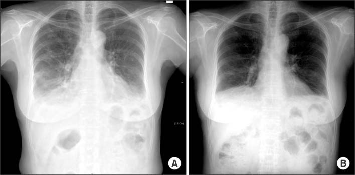
Figure 1 Chest X-ray on admission shows bilateral, multifocal consolidations and bilateral pleural effusion (A). After 3 months with steroid treatment, chest X-ray shows marked improvement (B).

Figure 2 High resolution computed tomography of chest on admission shows consolidation at right middle lobe (A) and multifocal ground grass opacity at both lower lobes (B). After 3 months with steroid treatment, marked improvement observed (C, D).
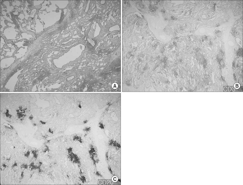
Figure 3 Histopathologic finding of the resected lung. Right lower lobe revealed lymphoid infilatration in the peribronchial, interstitial and alveolar septum (A, H&E stain, ×2). Immunohistochemical stains for CD3, CD20 revealed polyclonal lymphocytes (B, CD3 stain, ×2; C, CD20 stain, ×2).
Reference
-
1. Swigris JJ, Berry GJ, Raffin TA, Kuschner WG. Lymphoid interstitial pneumonia: a narrative review. Chest. 2002. 122:2150–2164.2. Carrington CB, Liebow AA. Lymphocytic interstitial pneumonia. Am J Pathol. 1966. 48:A36.3. Cosgrove G, Fessler M, Schwartz M. Schwarz MI, King TE, editors. Lymphocytoplasmic infiltrations of the lung. Interstitial lung disease. 2003. 4th ed. Hamilton: BC Decker;685–696.4. Yoo B, Kim NK, Kim KY, Han YC, Cho HI, Ham EK, et al. A case of lymphoma manifested as lymphocytic interstitial pneumonia. Korean J Intern Med. 1987. 33:386–392.5. Jung HJ, Cho ER, Shim JJ, In KH, Yu SH, Kang KH, et al. A case of lymphocytic interstitial pneumonitis. Tuberc Respir Dis. 1993. 40:602–609.6. Suh YA, Kim SI, Kim DH, Kwak JY, Lee JC, Baek HJ, et al. A case of lymphocytic interstitial pneumonia. Tuberc Respir Dis. 2001. 51:390–394.7. Yum HK, Kim ES, Ok KS, Lee HK, Choi SJ. Lymphocytic interstitial pneumonitis associated with epstein-barr virus in systemic lupus Erythematosus and Sjogren's syndrome: complete remission with corticosteroid and cyclophosphamide. Korean J Intern Med. 2002. 17:198–203.8. Scientific Committee of the Korean Academy of Tuberculosis and Respiratory Diseases. 2008 national survey of idiopathic interstitial pneumonia in Korea. Tuberc Respir Dis. 2009. 66:141–151.9. Johkoh T, Müller NL, Pickford HA, Hartman TE, Ichikado K, Akira M, et al. Lymphocytic interstitial pneumonia: thin-section CT findings in 22 patients. Radiology. 1999. 212:567–572.10. Honda O, Johkoh T, Ichikado K, Tomiyama N, Maeda M, Mihara N, et al. Differential diagnosis of lymphocytic interstitial pneumonia and malignant lymphoma on high-resolution CT. AJR Am J Roentgenol. 1999. 173:71–74.11. Koss MN, Hochholzer L, Langloss JM, Wehunt WD, Lazarus AA. Lymphoid interstitial pneumonia: clinicopathological and immunopathological findings in 18 cases. Pathology. 1987. 19:178–185.12. Strimlan CV, Rosenow EC 3rd, Weiland LH, Brown LR. Lymphocytic interstitial pneumonitis: review of 13 cases. Ann Intern Med. 1978. 88:616–621.13. Davies CW, Juniper MC, Gray W, Gleeson FV, Chapel HM, Davies RJ. Lymphoid interstitial pneumonitis associated with common variable hypogammaglobulinaemia treated with cyclosporin A. Thorax. 2000. 55:88–90.
- Full Text Links
-
- Actions
-
Cited
- CITED
-
- Close
- Share
-
- Similar articles
-
- Idiopathic Interstitial Pneumonias: Radiologic Findings
- Remission of Lymphocytic Interstitial Pneumonia in Sjogren's Syndrome after Autologous Peripheral Blood Stem Cell Transplantation
- Acute Interstitial Pneumonia: HRCT Findings in Five Patients
- Lymphocytic Interstitial Pneumonia in Primary Sjogren's Syndrome: A Case Report
- Extensive Bilateral Airspace Consolidation
