Consolidative Bronchioloalveolar Carcinoma Presenting as Pneumonia, and This Led to a Late Diagnosis due to the Improvement after Antibiotic Therapy
- Affiliations
-
- 1Department of Internal Medicine, Lung Institute of Medical Research Center, Seoul National University College of Medicine, Seoul, Department of Internal Medicine, Respiratory Center, Seoul National University Bundang Hospital, Seongnam, Korea. mdkang@medi
Abstract
- Non-resolving or slowly resolving pulmonary infiltrates in spite of administering adequate antimicrobial therapy are a clinical diagnostic challenge for physicians. The rate of radiographic resolution varies with the patients' age, the underlying comorbidities, the extent of radiographic involvement, the functional status and the causal pathogens. It is important to differentiate non-resolving or slowly resolving bacterial pneumonia from other uncommon infectious pneumonias or malignancies that require invasive diagnostic techniques to confirm the diagnosis. Bronchioloalveolar carcinoma can present with various clinical and radiographic features. Unfortunately, the radiographic similarity of consolidative BAC to pneumonia often leads to an incorrect diagnosis of pneumonia and possibly significant delays in obtaining appropriate diagnostic studies. We describe here a case of a mixed adenocarcinoma and bronchioloalveolar carcinoma that was initially diagnosed as pneumonia due to the consolidation pattern on the radiography and the patient's initial improvement with antibiotic treatment.
MeSH Terms
Figure
-
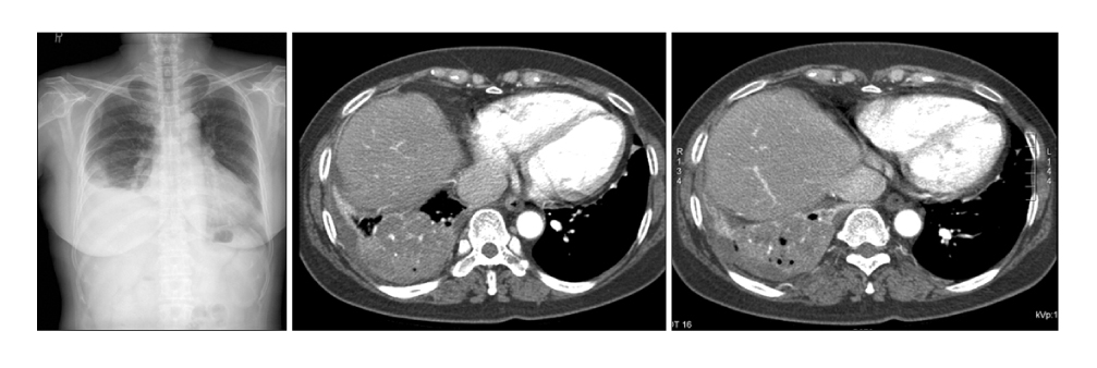
Figure 1 Initial chest CT (at admission) showed right lower lobe consolidation with pleural effusion and reactive LN enlargement.
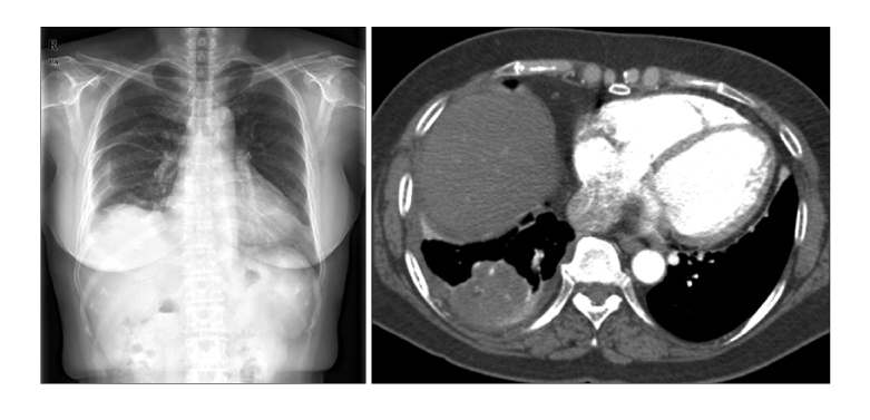
Figure 2 Chest images at 16th day after admission showed improved right lower lobe consolidation and pleural effusion after 16 days of antibiotics therapy.
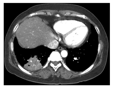
Figure 3 After 2 month of discharge, follow-up chest CT showed decreased pleural effusion but remaining consolidation was still observed at right lower lobe.
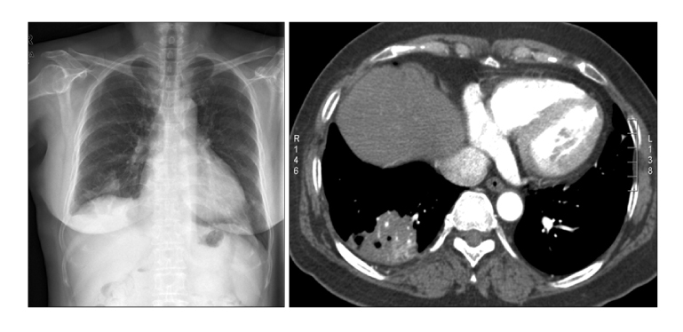
Figure 4 After 4 month of discharge, follow-up chest CT showed decreased but remaining consolidation with focal cavitation at right lower lobe.
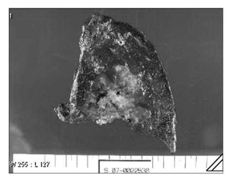
Figure 5 Resected lung tissue showed mass with necrosis and bleeding.
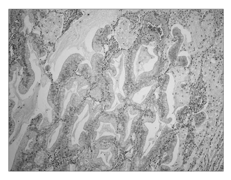
Figure 6 Tumor cells showed mucinous-bronchioloalveolar pattern composed of tall columnar cells with basal nuclei and pale cytoplasm, resembling goblet cells, with varying amounts of cytoplasmic mucin. It also showd extensive consolidation with numerous alveolar macrophages (H&E stain, ×100).
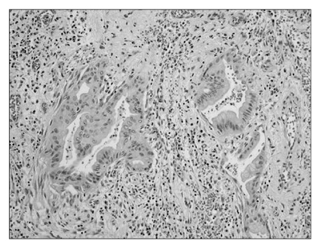
Figure 7 The resected lung specimen showed invasion which was characterized by significant increase in cytologic atypia, a fibroblastic stromal reaction, and an acinar pattern of growth (H&E stain, ×200).
Reference
-
1. Niederman MS. Understanding the natural history of community-acquired pneumonia resolution: vital information for optimizing duration of therapy. Clin Infect Dis. 2004. 39:1791–1793.2. Kuru T, Lynch JP 3rd. Nonresolving or slowly resolving pneumonia. Clin Chest Med. 1999. 20:623–651.3. El Solh AA, Aquilina AT, Gunen H, Ramadan F. Radiographic resolution of community-acquired bacterial pneumonia in the elderly. J Am Geriatr Soc. 2004. 52:224–229.4. Thompson WH. Bronchioloalveolar carcinoma masquerading as pneumonia. Respir Care. 2004. 49:1349–1353.5. Raz DJ, Kim JY, Jablons DM. Diagnosis and treatment of bronchioloalveolar carcinoma. Curr Opin Pulm Med. 2007. 13:290–296.6. Read WL, Page NC, Tierney RM, Piccirillo JF, Govindan R. The epidemiology of bronchioloalveolar carcinoma over the past two decades: analysis of the SEER database. Lung Cancer. 2004. 45:137–142.7. Sadohara J, Fujimoto K, Terasaki H, Nonoshita M, Hayabuchi N. Bronchioloalveolar carcinoma with fluctuating extent of consolidation on chest radiography. J Thorac Imaging. 2004. 19:63–66.8. Dumont P, Gasser B, Rouge C, Massard G, Wihlm JM. Bronchoalveolar carcinoma: histopathologic study of evolution in a series of 105 surgically treated patients. Chest. 1998. 113:391–395.9. Casey KR. Neoplastic mimics of pneumonia. Semin Respir Infect. 1995. 10:131–142.10. Aquino SL, Chiles C, Halford P. Distinction of consolidative bronchioloalveolar carcinoma from pneumonia: do CT criteria work? AJR Am J Roentgenol. 1998. 171:359–363.11. Kim MH, Kim CH, Kim YH, Park TW, Kang JH, Ahn MI, et al. A case of very slowly progressed pneumonic consolidation. Tuberc Respir Dis. 1996. 43:472–476.12. Kim TH, Kim SJ, Ryu YH, Chung SY, Seo JS, Kim YJ, et al. Differential CT features of infectious pneumonia versus bronchioloalveolar carcinoma (BAC) mimicking pneumonia. Eur Radiol. 2006. 16:1763–1768.
- Full Text Links
-
- Actions
-
Cited
- CITED
-
- Close
- Share
-
- Similar articles
-
- Diffe rentiation of Pneumonic-Type Bronchioloalveolar Carcinoma and Infectious Pneumonia in Clinical, Radiologic Findings
- Malignant Pulmonary Neoplasms Causing Airspace Consolidation: CT Findings
- CT Findings of Bronchioloalveolar Carcinoma
- A case of very slowly progressed pneumonic consolidation
- Computed tomography of bronchioloalveolar carcinoma showing open bronchus sign
