Restor Dent Endod.
2014 Aug;39(3):149-154.
In vitro cytotoxicity of four calcium silicate-based endodontic cements on human monocytes, a colorimetric MTT assay
- Affiliations
-
- 1Dental Research Center, Dentistry Research Institute, Tehran University of Medical Sciences, Tehran, Iran. nekoofarmh@cardiff.ac.uk
- 2Department of Endodontics, Dental School, Tehran University of Medical Sciences, Tehran, Iran.
- 3Department of Endodontics, Dental School, Qum University of Medical Sciences, Qum, Iran.
- 4Department of Immunology, Tehran University of Medical Sciences School of Medicine, Tehran, Iran.
- 5Endodontology Research Group, College of BioMedical and Life Sciences School of Dentistry, Cardiff University, Cardiff, UK.
Abstract
OBJECTIVES
This study was performed to evaluate the cytotoxicity of four calcium silicate-based endodontic cements at different storage times after mixing.
MATERIALS AND METHODS
Capillary tubes were filled with Biodentine (Septodont), Calcium Enriched Mixture (CEM cement, BioniqueDent), Tech Biosealer Endo (Tech Biosealer) and ProRoot MTA (Dentsply Tulsa Dental). Empty tubes and tubes containing Dycal were used as negative and positive control groups respectively. Filled capillary tubes were kept in 0.2 mL microtubes and incubated at 37degrees C. Each material was divided into 3 groups for testing at intervals of 24 hr, 7 day and 28 day after mixing. Human monocytes were isolated from peripheral blood mononuclear cells and cocultered with 24 hr, 7 day and 28 day samples of different materials for 24 and 48 hr. Cell viability was evaluated using an MTT assay.
RESULTS
In all groups, the viability of monocytes significantly improved with increasing storage time regardless of the incubation time (p < 0.001). After 24 hr of incubation, there was no significant difference between the materials regarding monocyte viability. However, at 48 hr of incubation, ProRoot MTA and Biodentine were less cytotoxic than CEM cement and Biosealer (p < 0.01).
CONCLUSIONS
Biodentine and ProRoot MTA had similar biocompatibility. Mixing ProRoot MTA with PBS in place of distilled water had no effect on its biocompatibility. Biosealer and CEM cement after 48 hr of incubation were significantly more cytotoxic to on monocyte cells compared to ProRoot MTA and Biodentine.
Keyword
Figure
-
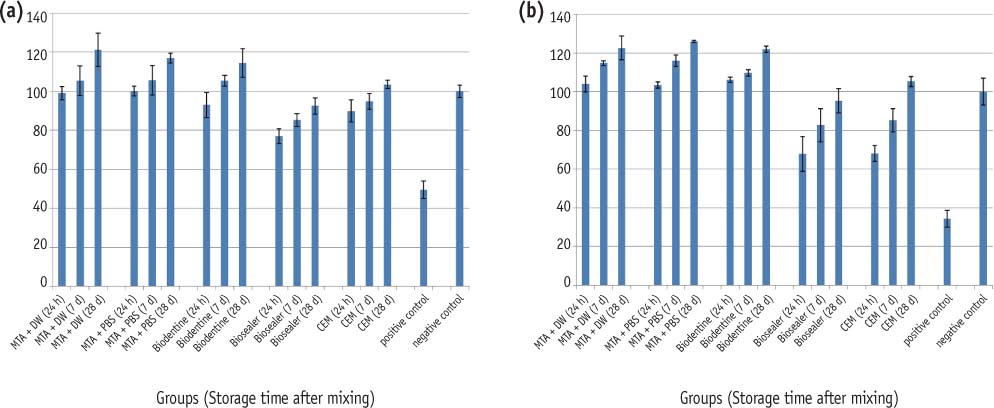
Figure 1 The metabolic activity of human monocytes in different experimental groups and storage times. (a) After 24 hours incubation time; (b) After 48 hours incubation time. MTA, Mineral trioxide aggregate; CEM, Calcium enriched mixture; DW, Distilled water; PBS, Phosphate buffered saline.
Reference
-
1. Parirokh M, Torabinejad M. Mineral trioxide aggregate: a comprehensive literature review-Part I: chemical, physical, and antibacterial properties. J Endod. 2010; 36:16–27.
Article2. Modareszadeh MR, Di Fiore PM, Tipton DA, Salamat N. Cytotoxicity and alkaline phosphatase activity evaluation of endosequence root repair material. J Endod. 2012; 38:1101–1105.
Article3. Ciasca M, Aminoshariae A, Jin G, Montagnese T, Mickel A. A comparison of the cytotoxicity and proinflammatory cytokine production of EndoSequence root repair material and ProRoot mineral trioxide aggregate in human osteoblast cell culture using reversetranscriptase polymerase chain reaction. J Endod. 2012; 38:486–489.
Article4. Torabinejad M, Parirokh M. Mineral trioxide aggregate: a comprehensive literature review-part II: leakage and biocompatibility investigations. J Endod. 2010; 36:190–202.
Article5. Hirschman WR, Wheater MA, Bringas JS, Hoen MM. Cytotoxicity comparison of three current direct pulp-capping agents with a new bioceramic root repair putty. J Endod. 2012; 38:385–388.
Article6. Chang SW, Yoo HM, Park DS, Oh TS, Bae KS. Ingredients and cytotoxicity of MTA and 3 kinds of Portland cements. J Korean Acad Conserv Dent. 2008; 33:369–376.
Article7. Villat C, Tran XV, Pradelle-Plasse N, Ponthiaux P, Wenger F, Grosgogeat B, Colon P. Impedance methodology: a new way to characterize the setting reaction of dental cements. Dent Mater. 2010; 26:1127–1132.
Article8. Laurent P, Camps J, De Méo M, Déjou J, About I. Induction of specific cell responses to a Ca(3)SiO(5)-based posterior restorative material. Dent Mater. 2008; 24:1486–1494.
Article9. Grech L, Mallia B, Camilleri J. Investigation of the physical properties of tricalcium silicate cement-based root-end filling materials. Dent Mater. 2013; 29:e20–e28.
Article10. Nosrat A, Seifi A, Asgary S. Regenerative endodontic treatment (revascularization) for necrotic immature permanent molars: a review and report of two cases with a new biomaterial. J Endod. 2011; 37:562–567.
Article11. Asgary S, Eghbal MJ, Parirokh M, Ghoddusi J, Kheirieh S, Brink F. Comparison of mineral trioxide aggregate's composition with Portland cements and a new endodontic cement. J Endod. 2009; 35:243–250.
Article12. Mozayeni MA, Milani AS, Marvasti LA, Asgary S. Cytotoxicity of calcium enriched mixture cement compared with mineral trioxide aggregate and intermediate restorative material. Aust Endod J. 2012; 38:70–75.
Article13. Ghoddusi J, Tavakkol Afshari J, Donyavi Z, Brook A, Disfani R, Esmaeelzadeh M. Cytotoxic effect of a new endodontic cement and mineral trioxide aggregate on L929 line culture. Iran Endod J. 2008; 3:17–23.14. Asgary S, Moosavi SH, Yadegari Z, Shahriari S. Cytotoxic effect of MTA and CEM cement in human gingival fibroblast cells. Scanning electronic microscope evaluation. N Y State Dent J. 2012; 78:51–54.15. Parirokh M, Mirsoltani B, Raoof M, Tabrizchi H, Haghdoost AA. Comparative study of subcutaneous tissue responses to a novel root-end filling material and white and grey mineral trioxide aggregate. Int Endod J. 2011; 44:283–289.
Article16. Rahimi S, Mokhtari H, Shahi S, Kazemi A, Asgary S, Eghbal MJ, Mesgariabbasi M, Mohajeri D. Osseous reaction to implantation of two endodontic cements: mineral trioxide aggregate (MTA) and calcium enriched mixture (CEM). Med Oral Patol Oral Cir Bucal. 2012; 17:e907–e911.
Article17. Gandolfi MG, Taddei P, Modena E, Siboni F, Prati C. Biointeractivity-related versus chemi/physisorptionrelated apatite precursor-forming ability of current root end filling materials. J Biomed Mater Res B Appl Biomater. 2013; 101:1107–1123.
Article18. Hakki S, Bozkurt B, Ozcopur B, Gandolfi MG, Prati C, Belli S. The response of cementoblasts to calcium phosphate resin-based and calcium silicate-based commercial sealers. Int Endod J. 2013; 46:242–252.
Article19. de Oliveira Mendes Oliveira, Ribeiro Sobrinho AP, de Carvalho AT, de Carvalho AT, de Souza Côrtes MI, Vieira LQ. In vitro evaluation of the cytotoxicity of two root canal sealers on macrophage activity. J Endod. 2003; 29:95–99.20. Vahid A, Hadjati J, Kermanshah H, Ghabraei S. Effects of cured dentin bonding materials on human monocyte viability. Oral Surg Oral Med Oral Pathol Oral Radiol Endod. 2004; 98:619–621.
Article21. Khedmat S, Hadjati J, Iravani A, Nourizadeh M. Effects of enamel matrix derivative on the viability, cytokine secretion, and phagocytic activity of human monocytes. J Endod. 2010; 36:1000–1003.
Article22. Gandolfi MG, Iacono F, Agee K, Siboni F, Tay F, Pashley DH, Prati C. Setting time and expansion in different soaking media of experimental accelerated calcium-silicate cements and ProRoot MTA. Oral Surg Oral Med Oral Pathol Oral Radiol Endod. 2009; 108:e39–e45.
Article23. Lotfi M, Vosoughhosseini S, Saghiri MA, Rahimi S, Zand V, Reyhani MF, Samiei M, Ghasemi N, Mehrvarzfar P, Azimi S, Shokohinejad N. Effect of synthetic tissue fluid on microleakage of grey and white mineral trioxide aggregate as root-end filling materials: an in vitro study. Sultan Qaboos Univ Med J. 2012; 12:323–329.
Article24. Asgary S, Eghbal MJ, Parirokh M, Ghoddusi J. Effect of two storage solutions on surface topography of two root-end fillings. Aust Endod J. 2009; 35:147–152.
Article25. Gandolfi MG, Taddei P, Tinti A, Prati C. Apatite-forming ability (bioactivity) of ProRoot MTA. Int Endod J. 2010; 43:917–929.
Article26. Han L, Okiji T. Bioactivity evaluation of three calcium silicate-based endodontic materials. Int Endod J. 2013; 46:808–814.
Article27. Shokouhinejad N, Nekoofar MH, Razmi H, Sajadi S, Davies TE, Saghiri MA, Gorjestani H, Dummer PM. Bioactivity of EndoSequence root repair material and bioaggregate. Int Endod J. 2012; 45:1127–1134.
Article28. Nekoofar MH, Adusei G, Sheykhrezae MS, Hayes SJ, Bryant ST, Dummer PM. The effect of condensation pressure on selected physical properties of mineral trioxide aggregate. Int Endod J. 2007; 40:453–461.
Article29. Nekoofar MH, Oloomi K, Sheykhrezae MS, Tabor R, Stone DF, Dummer PM. An evaluation of the effect of blood and human serum on the surface microhardness and surface microstructure of mineral trioxide aggregate. Int Endod J. 2010; 43:849–858.
Article30. Nekoofar MH, Aseeley Z, Dummer PM. The effect of various mixing techniques on the surface microhardness of mineral trioxide aggregate. Int Endod J. 2010; 43:312–320.
Article31. Asgary S, Shahabi S, Jafarzadeh T, Amini S, Kheirieh S. The properties of a new endodontic material. J Endod. 2008; 34:990–993.
Article32. Rezende TM, Vieira LQ, Cardoso FP, Oliveira RR, de Oliveira Mendes ST, Jorge ML, Ribeiro Sobrinho AP. The effect of mineral trioxide aggregate on phagocytic activity and production of reactive oxygen, nitrogen species and arginase activity by M1 and M2 macrophages. Int Endod J. 2007; 40:603–611.
Article33. Gomes-Filho JE, Watanabe S, Gomes AC, Faria MD, Lodi CS, Penha Oliveira SH. Evaluation of the effects of endodontic materials on fibroblast viability and cytokine production. J Endod. 2009; 35:1577–1579.
Article34. Kim HJ, Baek SH, Bae KS. Cytotoxicity and genotoxicity of newly developed calcium phosphate-based root canal sealers. J Korean Acad Conserv Dent. 2006; 31:36–49.
Article35. Lee JH, Baek SH, Bae KS. Evaluation of the cytotoxicity of calcium phosphate root canal sealers. J Korean Acad Conserv Dent. 2003; 28:295–302.
Article36. Parirokh M, Torabinejad M. Mineral trioxide aggregate: a comprehensive literature review-Part III: Clinical applications, drawbacks, and mechanism of action. J Endod. 2010; 36:400–413.
Article37. Han L, Okiji T, Okawa S. Morphological and chemical analysis of different precipitates on mineral trioxide aggregate immersed in different fluids. Dent Mater J. 2010; 29:512–517.
Article38. Oh MJ, Jeong YN, Bae IH, Yang SY, Park BJ, Koh JT, Hwang YC, Hwang IN, Oh WM. Biocompatibility of experimental mixture of mineral trioxide aggregate and glass ionomer cement. J Korean Acad Conserv Dent. 2010; 35:359–367.
Article39. Koubi G, Colon P, Franquin JC, Hartmann A, Richard G, Faure MO, Lambert G. Clinical evaluation of the performance and safety of a new dentine substitute, Biodentine, in the restoration of posterior teeth-a prospective study. Clin Oral Investig. 2013; 17:243–249.
Article40. Han L, Okiji T. Uptake of calcium and silicon released from calcium silicate-based endodontic materials into root canal dentine. Int Endod J. 2011; 44:1081–1087.
Article41. Zhou HM, Shen Y, Wang ZJ, Li L, Zheng YF, Häkkinen L, Haapasalo M. In vitro cytotoxicity evaluation of a novel root repair material. J Endod. 2013; 39:478–483.42. Pérard M, Le Clerc J, Watrin T, Meary F, Pérez F, Tricot-Doleux S, Pellen-Mussi P. Spheroid model study comparing the biocompatibility of Biodentine and MTA. J Mater Sci Mater Med. 2013; 24:1527–1534.
Article43. Asgary S, Shahabi S, Jafarzadeh T, Amini S, Kheirieh S. The properties of a new endodontic material. J Endod. 2008; 34:990–993.
Article44. Gandolfi MG, Baldi JV, Prati C. Biomaterials for pulpcapping: calcium-ions pulpflux through dentin in indirect pulp-capping. updated 2013 Oct 16. Available from: http://iadr.confex.com/iadr/2012rio/webprogram/Paper165368.html.
- Full Text Links
-
- Actions
-
Cited
- CITED
-
- Close
- Share
-
- Similar articles
-
- In vitro cytotoxicity of four kinds orthodontic band cements
- Calcium silicate-based root canal sealers: a literature review
- Surgical management of an accessory canal in a maxillary premolar: a case report
- Cytotoxicity of Various Calcium Silicate-based Materials with Stem Cells from Deciduous Teeth
- An in Vitro Study of the Effects of Different Dentin Bonding Agents on the Prevention of Tooth Discoloration and the Sealing Ability of Calcium Silicate-Based Cement in Regenerative Endodontic Procedures
