Use of ultrasound Doppler to determine tooth vitality in a discolored tooth after traumatic injury: its prospects and limitations
- Affiliations
-
- 1Department of Conservative Dentistry, Oral Science Research Center, Yonsei University College of Dentistry, Seoul, Korea. sunghopark@yuhs.ac
Abstract
- When a tooth shows discoloration and does not respond to the cold test or electric pulp test (EPT) after a traumatic injury, its diagnosis can be even more difficult due to the lack of proper diagnostic methods to evaluate its vitality. In these case reports, we hope to demonstrate that ultrasound Doppler might be successfully used to evaluate the vitality of the tooth after trauma, and help reduce unnecessary endodontic treatments. In all three of the present cases, the teeth were discolored after traumatic injuries and showed negative responses to the cold test and EPT. However, they showed distinctive vital reactions in the ultrasound Doppler test during the whole observation period. In the first case, the tooth color returned to normal, and the tooth showed a positive response to the cold test and EPT at 10 wk after the injury. In the second case, the tooth color had returned to its normal shade at 10 wk after the traumatic injury but remained insensitive to the cold test and EPT. In the third case, the discoloration was successfully treated with vital tooth bleaching.
Figure
-
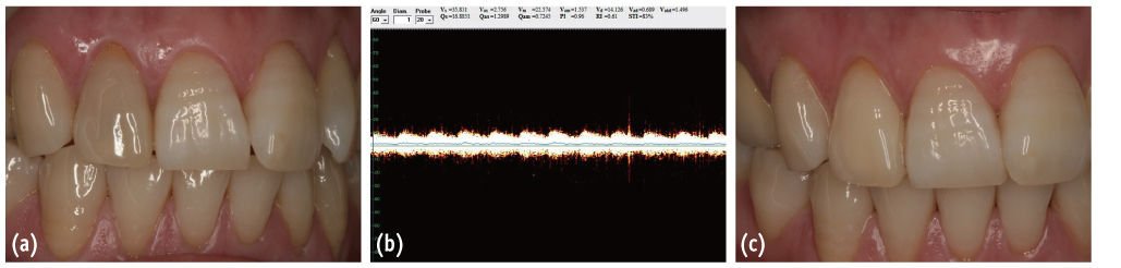
Figure 1 (a) In case 1, discoloration of tooth #12 was observed at 2 weeks after the injury; (b) The result of an ultrasound Doppler test at 6 weeks after the injury. It shows a typical pulsated image, which represents normal vital pulp; (c) At 10 weeks after the injury, the tooth had returned to a normal shade.
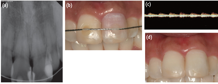
Figure 2 (a) In case 2, tooth #21 was splinted at a local clinic after a subluxation injury that had occurred 2 weeks before the patient visited our clinic. It showed a negative response to the thermal test and EPT, and a positive response to the percussion test; (b) Tooth #21 showed pinkish discoloration; (c) In the ultrasound Doppler test, tooth #21 showed a normal pulsated response like that of the other teeth; (d) At 10 weeks after the injury, the shade of tooth #21 had returned to normal.
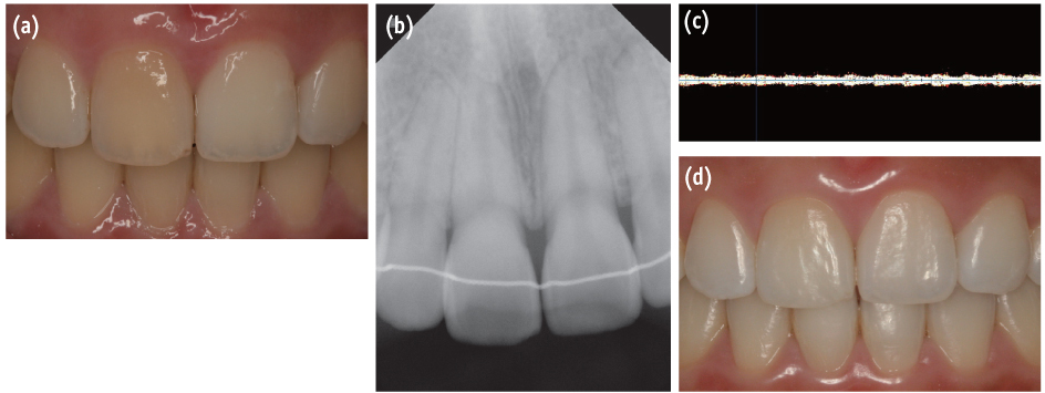
Figure 3 (a) In case 3, tooth #11 showed yellowish brown discoloration; (b) The coronal pulp space was obliterated, whereas the pulp space was present in the root area. There was no radiolucency in the periapical area, but the root apex was slightly shortened; (c) In the ultrasound Doppler test, tooth #11 showed an image typical of a vital tooth; (d) The patient was satisfied with the shade of tooth #11 after vital bleaching treatment.
Reference
-
1. Yoon MJ, Kim E, Lee SJ, Bae YM, Kim S, Park SH. Pulpal blood flow measurement with ultrasound Doppler imaging. J Endod. 2010; 36:419–422.
Article2. Abd-Elmeguid A, Yu DC. Dental pulp neurophysiology: part 2. Current diagnostic tests to assess pulp vitality. J Can Dent Assoc. 2009; 75:139–143.3. Ozçelik B, Kuraner T, Kendir B, Aşan E. Histopathological evaluation of the dental pulps in crown-fractured teeth. J Endod. 2000; 26:271–273.
Article4. Aguiló L, Gandía JL. Transient red discoloration: report of case. ASDC J Dent Child. 1998; 65:346–348. 3565. Andreasen FM. Pulpal healing after luxation injuries and root fracture in the permanent dentition. Endod Dent Traumatol. 1989; 5:111–131.
Article6. Malmgren B, Hübel S. Transient discoloration of the coronal fragment in intra-alveolar root fractures. Dent Traumatol. 2012; 28:200–204.
Article7. Andreasen FM. Transient apical breakdown and its relation to color and sensibility changes after luxation injuries to teeth. Endod Dent Traumatol. 1986; 2:9–19.
Article8. Cohenca N, Karni S, Rotstein I. Transient apical breakdown following tooth luxation. Dent Traumatol. 2003; 19:289–291.
Article9. Cotti E, Campisi G, Ambu R, Dettori C. Ultrasound real-time imaging in the differential diagnosis of periapical lesions. Int Endod J. 2003; 36:556–563.
Article10. Rajendran N, Sundaresan B. Efficacy of ultrasound and color power Doppler as a monitoring tool in the healing of endodontic periapical lesions. J Endod. 2007; 33:181–186.
Article11. Lustig JP, London D, Dor BL, Yanko R. Ultrasound identification and quantitative measurement of blood supply to the anterior part of the mandible. Oral Surg Oral Med Oral Pathol Oral Radiol Endod. 2003; 96:625–629.
Article12. Yoon MJ, Lee SJ, Kim E, Park SH. Doppler ultrasound to detect pulpal blood flow changes during local anaesthesia. Int Endod J. 2012; 45:83–87.
Article13. Cohen S, Hargreaves KM. Pathways of the pulp. 9th ed. Louis: Mosby;2006. p. 504–508.14. Ehrmann EH. Pulp testers and pulp testing with particular reference to the use of dry ice. Aust Dent J. 1977; 22:272–279.
Article15. Klein H. Pulp responses to an electric pulp stimulator in the developing permanent anterior dentition. ASDC J Dent Child. 1978; 45:199–202.16. Olgart L, Gazelius B, Lindh-Strömberg U. Laser Doppler flowmetry in assessing vitality in luxated permanent teeth. Int Endod J. 1988; 21:300–306.
Article17. Sasano T, Onodera D, Hashimoto K, Iikubo M, Satoh-Kuriwada S, Shoji N, Miyahara T. Possible application of transmitted laser light for the assessment of human pulp vitality. Part 2. Increased laser power for enhanced detection of pulpal blood flow. Dent Traumatol. 2005; 21:37–41.
Article18. Gopikrishna V, Tinagupta K, Kandaswamy D. Comparison of electrical, thermal, and pulse oximetry methods for assessing pulp vitality in recently traumatized teeth. J Endod. 2007; 33:531–535.
Article19. Gopikrishna V, Tinagupta K, Kandaswamy D. Evaluation of efficacy of a new custom-made pulse oximeter dental probe in comparison with the electrical and thermal tests for assessing pulp vitality. J Endod. 2007; 33:411–414.
Article20. Jafarzadeh H, Rosenberg PA. Pulse oximetry: review of a potential aid in endodontic diagnosis. J Endod. 2009; 35:329–333.
Article21. Heithersay GS, Hirsch RS. Tooth discoloration and resolution following a luxation injury: significance of blood pigment in dentin to laser Doppler flowmetry readings. Quintessence Int. 1993; 24:669–676.
- Full Text Links
-
- Actions
-
Cited
- CITED
-
- Close
- Share
-
- Similar articles
-
- Changes in pulpal blood flow during orthodontic tooth movement studied by Doppler ultrasound
- Pulp vitality and coronal discoloration following traumatic injuries
- Autotransplantation combined with orthodontic treatment: a case involving the maxillary central incisors with root resorption after traumatic injury
- Diagnosis of periapical cemental dysplasia
- THE PROGNOSIS OF THE TEETH IN THE MANDIBULAR FRACTURE LINES
