J Korean Acad Conserv Dent.
2010 Nov;35(6):492-496. 10.5395/JKACD.2010.35.6.492.
Pulp vitality and coronal discoloration following traumatic injuries
- Affiliations
-
- 1Microscope Center, Department of Conservative Dentistry, Yonsei University, Seoul, Korea. andyendo@yuhs.ac
- KMID: 2176472
- DOI: http://doi.org/10.5395/JKACD.2010.35.6.492
Abstract
- Coronal discoloration is a common sequela to traumatic injuries. In subluxation cases, although the injury is not strong enough to rupture the apical vessels, discoloration may appear by tearing thin walls or occluding small capillaries. In absence of infection pulpal regeneration can occur, and as a result discoloration may completely or partially subside. But judging pulpal status by coronal discoloration can be dangerous and it may lead to unnecessary treatment. This case presents coronal discoloration and recovery following traumatic injury of maxillary anterior teeth. In diagnosing traumatized teeth routine cold tests or electric pulp tests are known to be unreliable, but with the aid of ultrasound doppler imaging, assessing pulp vitality of traumatized teeth can be more accurate.
Keyword
Figure
-

Figure 1 Periapical view at the first exam and after tooth reduction.
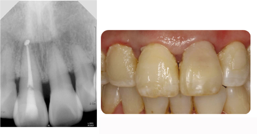
Figure 2 Periapical view and clinical photo at 2 weeks follow-up (discoloration on #21).
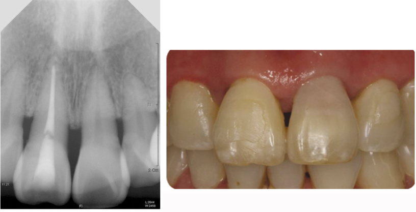
Figure 3 Periapical view and clinical photo at 6 weeks follow-up (discoloration on #21).

Figure 4 Ultrasound doppler graphy imaging on #11, 21, 22 (from top).

Figure 5 Periapical view and clinical photo at 6 weeks follow-up (regain the color on #21).
Cited by 1 articles
-
Pulp necrosis following luxated injury to teeth in a patient with uncontrolled type II diabetes mellitus: a case report
Haneol Shin, Seung-Jong Lee, Il-Young Jung, Chan-Young Lee
Restor Dent Endod. 2012;37(1):61-65. doi: 10.5395/rde.2012.37.1.61.
Reference
-
1. Andreasen FM, Vestergaard Pedersen B. Prognosis of luxated permanent teeth: the development of pulp necrosis. Endod Dent Traumatol. 1985. 1(6):207–220.2. Yu DC, Abd-Elmeguid A. Dental pulp neurophysiology: Part 2. Current diagnostic tests to assess pulp vitality. J Can Dent Assoc. 2009. 75(2):139–143.3. Aguiló L, Gandía JL. Transient red discoloration: report of case. ASDC J Dent Child. 1998. Sep-Oct. 65(5):346–348. 3564. Yoon MJ, Kim E, Lee SJ, Bae YM, Kim S, Park SH. Pulpal blood flow measurement with ultrasound Doppler imaging. J Endod. 2010. 36(3):419–422.
Article5. Andreasen JO. Textbook and color atlas of Traumatic injuries to the teeth. 4th ed. UK: Oxford.6. Ozçelik B, Kuraner T, Kendir B, Aşan E. Histopathological evaluation of the dental pulps in crown-fractured teeth. J Endod. 2000. 26(5):271–273.
Article7. Andreasen FM. Pulpal healing after luxation injuries and root fracture in the permanent dentition. Endod Dent Traumatol. 1989. 5(3):111–131.
Article8. Cohen S, et al. Pathways of the pulp. 9th ed. St. Louis: Mosby.9. Petersson K, Söderström C, Kiani-Anaraki M, Lévy G. Evaluation of the ability of thermal and electrical tests to register pulp vitality. Endod Dent Traumatol. 1999. 15(3):127–131.
Article10. Berson M, Grégoire JM, Gens F, Rateau J, Jamet F, Vaillant L, Tranquart F, Pourcelot L. High frequency(20MHz) ultrasonic devices : advantages and applications. Eur J Ultrasound. 1999. 10(1):53–63.
- Full Text Links
-
- Actions
-
Cited
- CITED
-
- Close
- Share
-
- Similar articles
-
- Use of ultrasound Doppler to determine tooth vitality in a discolored tooth after traumatic injury: its prospects and limitations
- Treatment of horizontal root-fractured maxillary incisors
- Coronal tooth discoloration induced by regenerative endodontic treatment using different scaffolds and intracanal coronal barriers: a 6-month ex vivo study
- Prevention of tooth discoloration associated with triple antibiotics
- Impaction of permanent canine caused by unsuccessful primary canine pulpotomy: a case report
