Obstet Gynecol Sci.
2015 Nov;58(6):446-452. 10.5468/ogs.2015.58.6.446.
The practice patterns of second trimester fetal ultrasonography: A questionnaire survey and an analysis of checklists
- Affiliations
-
- 1Department of Obstetrics and Gynecology, Graduate School of Medicine, Dongguk University, Seoul, Korea.
- 2Department of Obstetrics and Gynecology, Kyungpook National University School of Medicine, Daegu, Korea. wjseong@knu.ac.kr
- 3Department of Obstetrics and Gynecology, Seoul National University Bundang Hospital, Seongnam, Korea.
- 4Department of Obstetrics and Gynecology, Kyung Hee University School of Medicine, Seoul, Korea.
- 5Division of Maternal and Fetal Medicine, Department of Obstetrics and Gynecology, Konkuk University Medical Center, Konkuk University School of Medicine, Seoul, Korea.
- 6Hamchoon Women's Clinic, Seoul, Korea.
- 7Department of Obstetrics and Gynecology, Seoul St. Mary's Hospital, The Catholic University of Korea College of Medicine, Seoul, Korea.
- 8Department of Obstetrics and Gynecology, Cheil General Hospital and Women's Healthcare Center, Dankook University College of Medicine, Seoul, Korea.
- 9Department of Obstetrics and Gynecology, Samsung Medical Center, Sungkyunkwan University School of Medicine, Seoul, Korea.
- 10Department of Obstetrics and Gynecology, Bucheon St. Mary's Hospital, The Catholic University of Korea College of Medicine, Bucheon, Korea.
- KMID: 2314059
- DOI: http://doi.org/10.5468/ogs.2015.58.6.446
Abstract
OBJECTIVE
To analyze practice patterns and checklists of second trimester ultrasonography, and to investigate management plans when soft markers are detected among Korean Society of Ultrasound in Obstetrics and Gynecology (KSUOG) members.
METHODS
An internet-based self-administered questionnaire survey was designed. KSUOG members were invited to the survey. Checklists of the second trimester ultrasonography were also requested. In the questionnaire survey, general practice patterns of the second trimester ultrasonography and management schemes of soft markers were asked. In the checklists analysis, the number of items were counted and also compared with those recommended by other medical societies.
RESULTS
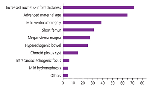
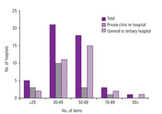
A total of 101 members responded. Eighty-seven percent routinely recommended second trimester fetal anatomic surveillance. Most (91.1%) performed it between 20+0 and 23+6 weeks of gestation. Written informed consents were given by 15.8% of respondents. Nearly 60% recommended genetic counseling when multiple soft markers and/or advanced maternal age were found. Similar tendencies were found in the managements of individual soft markers. However, practice patterns were very diverse and sometimes conflicting. Forty-eight checklists were analyzed in context with the number and content of the items. The median item number was 46.5 (range, 17 to 109). Of 49 items of checklists recommended by International Society of Ultrasound in Obstetrics and Gynecology and/or American Congress of Obstetricians and Gynecologists, 14 items (28.6%) were found in less than 50% of the checklists analyzed in this study.
CONCLUSION
Although general practice patterns were similar among KSUOG members, some of which were conflicting, and there is a need for standardization of the practice patterns and checklists of second trimester ultrasonography, which also have very wide range of spectrum.
Keyword
MeSH Terms
Figure
Reference
-
1. American College of Radiology. ACR-ACOG-AIUM-SRU practice guideline for the performance of obstetrical ultrasound: amended 2014 (resolution 39) [Internet]. Reston (VA): American College of Radiology;2014. cited 2015 Mar 1. Available from: http://www.acr.org/%7E/media/F7BC35BD59264E7CBE648F6D1BB8B8E2.pdf.2. Salomon LJ, Alfirevic Z, Berghella V, Bilardo C, Hernandez-Andrade E, Johnsen SL, et al. Practice guidelines for performance of the routine mid-trimester fetal ultrasound scan. Ultrasound Obstet Gynecol. 2011; 37:116–126.3. Bethune M. Literature review and suggested protocol for managing ultrasound soft markers for Down syndrome: thickened nuchal fold, echogenic bowel, shortened femur, shortened humerus, pyelectasis and absent or hypoplastic nasal bone. Australas Radiol. 2007; 51:218–225.4. Breathnach FM, Fleming A, Malone FD. The second trimester genetic sonogram. Am J Med Genet C Semin Med Genet. 2007; 145C:62–72.5. Agathokleous M, Chaveeva P, Poon LC, Kosinski P, Nicolaides KH. Meta-analysis of second-trimester markers for trisomy 21. Ultrasound Obstet Gynecol. 2013; 41:247–261.6. Raniga S, Desai PD, Parikh H. Ultrasonographic soft markers of aneuploidy in second trimester: are we lost? MedGenMed. 2006; 8:9.7. Rebarber A, Levey KA, Funai E, Monda S, Paidas M. An ethnic predilection for fetal echogenic intracardiac focus identified during targeted midtrimester ultrasound examination: a retrospective review. BMC Pregnancy Childbirth. 2004; 4:12.8. Shipp TD, Bromley B, Lieberman E, Benacerraf BR. The frequency of the detection of fetal echogenic intracardiac foci with respect to maternal race. Ultrasound Obstet Gynecol. 2000; 15:460–462.9. Bronsteen R, Lee W, Vettraino IM, Huang R, Comstock CH. Second-trimester sonography and trisomy 18: the significance of isolated choroid plexus cysts after an examination that includes the fetal hands. J Ultrasound Med. 2004; 23:241–245.10. Coco C, Jeanty P. Karyotyping of fetuses with isolated choroid plexus cysts is not justified in an unselected population. J Ultrasound Med. 2004; 23:899–906.11. Demasio K, Canterino J, Ananth C, Fernandez C, Smulian J, Vintzileos A. Isolated choroid plexus cyst in low-risk women less than 35 years old. Am J Obstet Gynecol. 2002; 187:1246–1249.12. Kim KW, Kwak DW, Ko HS, Park HS, Seol HJ, Hong JS, et al. The clinical practice patterns of fetal ultrasonography in the first-trimester: a questionnaire survey of members of the Korean Society of Ultrasound in Obstetrics and Gynecology. Obstet Gynecol Sci. 2014; 57:448–456.
- Full Text Links
- Actions
-
Cited
- CITED
-
- Close
- Share
- Similar articles
-
- The clinical practice patterns of fetal ultrasonography in the first-trimester: A questionnaire survey of members of the Korean Society of Ultrasound in Obstetrics and Gynecology
- 2014 First-trimester ultrasound forum from the Korean Society of Ultrasound in Obstetrics and Gynecology
- A Case of Fetal Cholelithiasis Related to Maternal Intrahepatic Cholestasis of Pregnancy
- A case of fetal intracranial hemorrhage diagnosed by antenatal ultrasonographic examination
- Prenatal ultrasonography of craniofacial abnormalities


