Obstet Gynecol Sci.
2014 Nov;57(6):448-456. 10.5468/ogs.2014.57.6.448.
The clinical practice patterns of fetal ultrasonography in the first-trimester: A questionnaire survey of members of the Korean Society of Ultrasound in Obstetrics and Gynecology
- Affiliations
-
- 1Department of Obstetrics and Gynecology, Hamchoon Women's Clinic, Seoul, Korea.
- 2Department of Obstetrics and Gynecology, Cheil General Hospital & Women's Healthcare Center, Kwandong University College of Medicine, Seoul, Korea.
- 3Department of Obstetrics and Gynecology, The Catholic University College of Medicine, Seoul, Korea.
- 4Department of Obstetrics and Gynecology, Dongguk University Ilsan Hospital, Goyang, Korea.
- 5Department of Obstetrics and Gynecology, Kyung Hee University Hospital, Seoul, Korea.
- 6Department of Obstetrics and Gynecology, Seoul National University Bundang Hospital, Seongnam, Korea.
- 7Department of Obstetrics and Gynecology, Konkuk University Medical Center, Konkuk University School of Medicine, Seoul, Korea. hwanghs@kuh.ac.kr
- 8Department of Obstetrics and Gynecology, Samsung Medical Center, Sungkyunkwan University School of Medicine, Seoul, Korea.
- KMID: 2314010
- DOI: http://doi.org/10.5468/ogs.2014.57.6.448
Abstract
OBJECTIVE
This study aimed to survey the current clinical practice of first-trimester ultrasonography among members of the Korean Society of Ultrasound in Obstetrics and Gynecology (KSUOG) and to provide basic data for making practical recommendations about first-trimester ultrasonography scan in Korea.
METHODS
This survey was conducted using a self-administered anonymous questionnaire. The first-trimester in this survey was divided into two parts: early and late first-trimester. The survey was focused on safety issue, nuchal translucency (NT) cutoff, the anatomic structures they check, and the need for practical recommendations or educational courses during the first-trimester.
RESULTS
During the study period, 194 KSUOG members participated into this survey. The survey on early first-trimester scan reveal that 173 (89.2%) of respondents had used pulsed-wave Doppler or color Doppler imaging to monitor fetal heart beat. For the late first-trimester scan, 145 (74.7%) of respondents was found to check for fetal anatomical assessments during their NT screening performance; however, the clinical practice patterns were considerably varied among participants. More than half of the respondents used the criterion of NT > or =3.0 mm to define increased NT. Approximately 80% of respondents stated that the screening ultrasonography of fetal structures in the first-trimester was necessary. Furthermore, 187 (96.4%) of respondents were in favor of a recommendation for first-trimester ultrasonography in Korea.
CONCLUSION
This is the first survey of the current clinical practice of first-trimester ultrasonography in Korea. Our survey findings highlight the need for the practical recommendation or educational course for first-trimester ultrasonography.
MeSH Terms
Figure
-
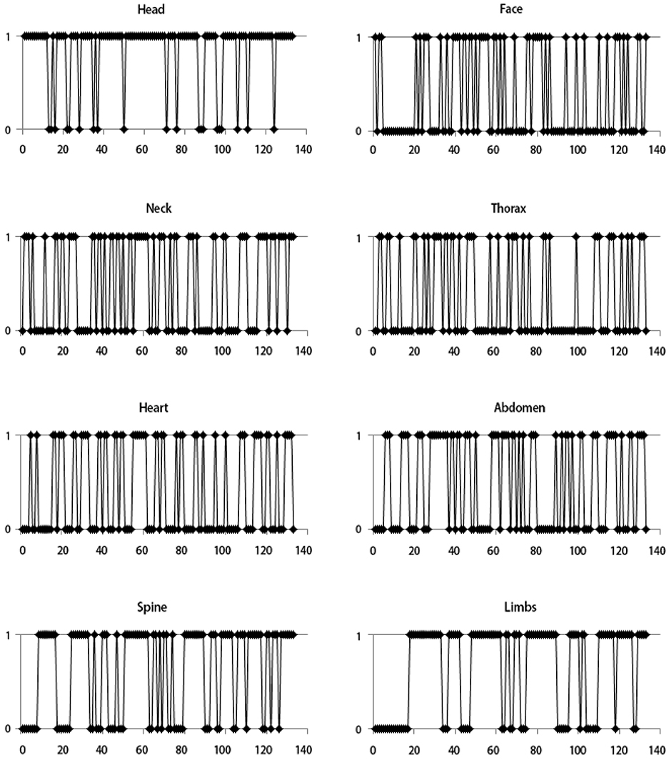
Fig. 1 Ultrasonographic tracings for eight fetal structural parts during the late first-trimester. The pattern of tracings for the eight parts (head, face, neck, thorax, heart, abdomen, spine, and limbs) differed significantly among respondents. X axis means individual respondents and Y axis means answers for checking the parts; 1 means checking and 0 means not checking.
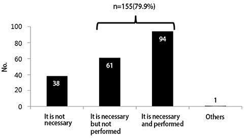
Fig. 2 The need for late first-trimester ultrasonography to detect fetal structural abnormalities; 79.9% considered it necessary.
Reference
-
1. McNay MB, Fleming JE. Forty years of obstetric ultrasonography 1957-1997: from A-scope to three dimensions. Ultrasound Med Biol. 1999; 25:3–56.2. Grant SS. Options for Down syndrome screening: what will women choose? J Midwifery Womens Health. 2005; 50:211–218.3. Chen M, Lee CP, Lam YH, Tang RY, Chan BC, Wong SF, et al. Comparison of nuchal and detailed morphology ultrasonography examinations in early pregnancy for fetal structural abnormality screening: a randomized controlled trial. Ultrasound Obstet Gynecol. 2008; 31:136–146.4. Saltvedt S, Almström H, Kublickas M, Valentin L, Grunewald C. Detection of malformations in chromosomally normal fetuses by routine ultrasonography at 12 or 18 weeks of gestation-a randomised controlled trial in 39,572 pregnancies. BJOG. 2006; 113:664–674.5. Timor-Tritsch IE, Fuchs KM, Monteagudo A, D'alton ME. Performing a fetal anatomy scan at the time of first-trimester screening. Obstet Gynecol. 2009; 113:402–407.6. Cedergren M, Selbing A. Detection of fetal structural abnormalities by an 11-14-week ultrasonography dating scan in an unselected Swedish population. Acta Obstet Gynecol Scand. 2006; 85:912–915.7. Taipale P, Ammala M, Salonen R, Hiilesmaa V. Learning curve in ultrasonographic screening for selected fetal structural anomalies in early pregnancy. Obstet Gynecol. 2003; 101:273–278.8. Fisher J. First-trimester screening: dealing with the fall-out. Prenat Diagn. 2011; 31:46–49.9. Rossi AC, Prefumo F. Accuracy of ultrasonography at 11-14 weeks of gestation for detection of fetal structural anomalies: a systematic review. Obstet Gynecol. 2013; 122:1160–1167.10. Salomon LJ, Alfirevic Z, Bilardo CM, Chalouhi GE, Ghi T, Kagan KO, et al. ISUOG practice guidelines: performance of first-trimester fetal ultrasonography scan. Ultrasound Obstet Gynecol. 2013; 41:102–113.11. Hershkovitz R, Sheiner E, Mazor M. Ultrasonography in obstetrics: a review of safety. Eur J Obstet Gynecol Reprod Biol. 2002; 101:15–18.12. Salvesen K, Lees C, Abramowicz J, Brezinka C, Ter Haar G, Marsal K, et al. ISUOG statement on the safe use of Doppler in the 11 to 13 +6-week fetal ultrasonography examination. Ultrasound Obstet Gynecol. 2011; 37:628.13. Snijders RJ, Noble P, Sebire N, Souka A, Nicolaides KH. Fetal Medicine Foundation First Trimester Screening Group. UK multicentre project on assessment of risk of trisomy 21 by maternal age and fetal nuchal-translucency thickness at 10-14 weeks of gestation. Lancet. 1998; 352:343–346.14. Wapner R, Thom E, Simpson JL, Pergament E, Silver R, Filkins K, et al. First-trimester screening for trisomies 21 and 18. N Engl J Med. 2003; 349:1405–1413.15. Malone FD, Canick JA, Ball RH, Nyberg DA, Comstock CH, Bukowski R, et al. First-trimester or second-trimester screening, or both, for Down's syndrome. N Engl J Med. 2005; 353:2001–2011.16. Nicolaides KH, Azar G, Byrne D, Mansur C, Marks K. Fetal nuchal translucency: ultrasonography screening for chromosomal defects in first trimester of pregnancy. BMJ. 1992; 304:867–869.17. Wald NJ, Rodeck C, Hackshaw AK, Walters J, Chitty L, Mackinson AM. First and second trimester antenatal screening for Down's syndrome: the results of the Serum, Urine and ultrasonography Screening Study (SURUSS). J Med Screen. 2003; 10(2):56–104.18. Nicolaides KH, Spencer K, Avgidou K, Faiola S, Falcon O. Multicenter study of first-trimester screening for trisomy 21 in 75 821 pregnancies: results and estimation of the potential impact of individual risk-orientated two-stage first-trimester screening. Ultrasound Obstet Gynecol. 2005; 25:221–226.19. Jouannic JM, Thieulin AC, Bonnet D, Houyel L, Lelong N, Goffinet F, et al. Measurement of nuchal translucency for prenatal screening of congenital heart defects: a population-based evaluation. Prenat Diagn. 2011; 31:1264–1269.20. Simpson LL, Malone FD, Bianchi DW, Ball RH, Nyberg DA, Comstock CH, et al. Nuchal translucency and the risk of congenital heart disease. Obstet Gynecol. 2007; 109:376–383.21. Souka AP, Von Kaisenberg CS, Hyett JA, Sonek JD, Nicolaides KH. Increased nuchal translucency with normal karyotype. Am J Obstet Gynecol. 2005; 192:1005–1021.22. Cuckle H. Monitoring quality control of nuchal translucency. Clin Lab Med. 2010; 30:593–604.23. Sahota DS, Leung WC, To WK, Chan WP, Lau TK, Leung TY. Quality assurance of nuchal translucency for prenatal fetal Down syndrome screening. J Matern Fetal Neonatal Med. 2012; 25:1039–1043.24. D'Alton ME. Nuchal translucency quality monitoring: the transition from research to clinical care. Obstet Gynecol. 2010; 116:806–807.
- Full Text Links
-
- Actions
-
Cited
- CITED
-
- Close
- Share
-
- Similar articles
-
- The practice patterns of second trimester fetal ultrasonography: A questionnaire survey and an analysis of checklists
- 2014 First-trimester ultrasound forum from the Korean Society of Ultrasound in Obstetrics and Gynecology
- The Value of Ultrasonography in Obstetrics and Gynecology
- Fetal Wastage in the Second-Trimester of Pregnancy: An Obstetric Approach
- Management of isolated oligohydramnios in Korea: a questionnaire-based study of clinical practice patterns among the members of the Korean Society of Maternal Fetal Medicine
