Lab Med Online.
2012 Jul;2(3):179-180.
Acanthocytosis in a Patient with Chorea-acanthocytosis
- Affiliations
-
- 1Department of Laboratory Medicine, Pusan National University Yangsan Hospital, Yangsan, Korea.
- 2Department of Neurology, Pusan National University Yangsan Hospital, Yangsan, Korea. drleejae@hanmail.net
- 3Research Institute for Convergence of Biomedical Science and Technology, Pusan National University Yangsan Hospital, Yangsan, Korea.
Abstract
- No abstract available.
Figure
-
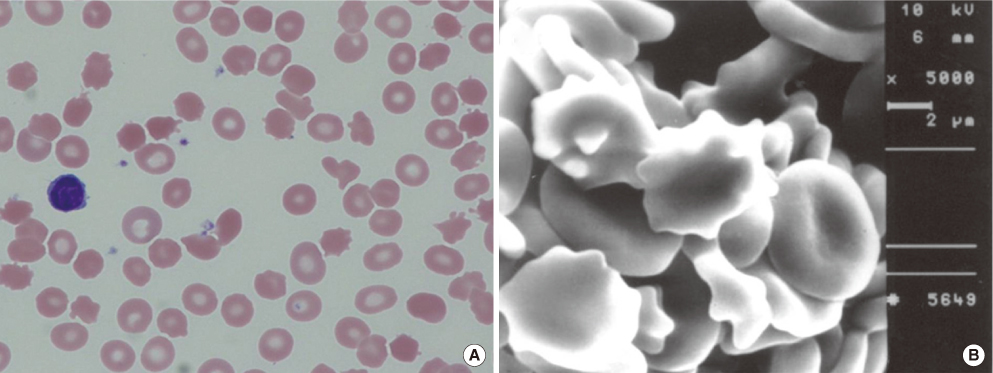
Fig. 1 Peripheral blood smear demonstrates many acanthocytes that do not have central pallor and are smaller than normocytic red blood cells (A) (Wright stain,×1,000). Scanning electron microscopy discloses many acanthocytes characterized by their thorn-like protrusions (B) (SEM,×1,000).
Reference
-
1. Walker RH, Jung HH, Dobson-Stone C, Rampoldi L, Sano A, Tison F, et al. Neurologic phenotypes associated with acanthocytosis. Neurology. 2007. 68:92–98.
Article2. Storch A, Kornhass M, Schwarz J. Testing for acanthocytosis, A prospective reader-blinded study in movement disorder patients. J Neurol. 2005. 252:84–90.
- Full Text Links
-
- Actions
-
Cited
- CITED
-
- Close
- Share
-
- Similar articles
-
- Globus Pallidus Interna Deep Brain Stimulation for Chorea-Acanthocytosis
- Treatment of Psychiatric Symptoms in a Patient with Neuroacanthocytosis: A Case Report
- Neuroacanthocytosis Syndrome Misdiagnosed as Inflammatory Myopathy: A Case Report
- Treatment of a lip defect in a patient with chorea-acanthocytosis using a combination of surgical and adjuvant onabotulinumtoxinA therapy: a case report
- Pallidus Stimulation for Chorea-Acanthocytosis: A Systematic Review and Meta-Analysis of Individual Data
