Lab Med Online.
2011 Jul;1(3):168-171.
A Case of Masked t(8;21) in Acute Myeloid Leukemia with Cytogenetic Abnormality of 45,X,-Y,t(8;17)(q22;p13)
- Affiliations
-
- 1Department of Laboratory Medicine, Wonkwang University School of Medicine, Iksan, Korea. jin20@wku.ac.kr
- 2Department of Internal Medicine, Wonkwang University School of Medicine, Iksan, Korea.
- 3Institute of Wonkwang Medical Science, Wonkwang University School of Medicine, Iksan, Korea.
Abstract
- The t(8;21)(q22;q22) is one of the most frequent structural chromosomal anomaly found in AML, occurring in about 5% of all AML and in 10% of AML with maturation (M2). And approximately 3.4% of AML with t(8;21)(q22;q22) occurs as a complex chromosomal abnormality and occasionally shows discrepancy between cytogenetic and molecular genetic analyses. We report a case of 42 yr old male patient that revealed morphological characteristics of AML-M2 and karyotypic abnormality of 45,X,-Y,t(8;17)(q22;p13) without visible involvement of chromosome 21 by conventional cytogenetic study with masked t(8;21) identified by FISH using RUNX1/RUNX1T1 probes. FISH confirmed nuc ish (RUNX1T1x3),(RUNX1x3), (RUNX1T1 con RUNX1x1). According to the results of conventional cytogenetic and FISH analyses, the karyotype was revised to 45,X,-Y,t(8;17;21)(q22;p13;q22).
Keyword
MeSH Terms
Figure
-
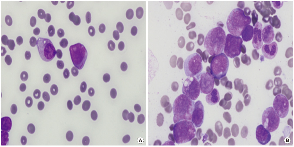
Fig. 1 The blasts in (A) peripheral blood smear containing single Auer rod and in (B) bone marrow aspirate smear (Wright-Giemsa stain, ×1,000). They reveal large sized, round shaped nuclei with prominent nucleoli and moderate to abundant cytoplasm with azurophilic granules.
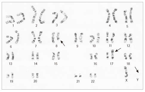
Fig. 2 The G-banded karyotype of the metaphases analyzed in the patient's bone marrow cells shows 45,X,-Y,t(8;17)(q22;p13). Arrows indicate abnormal chromosomes.
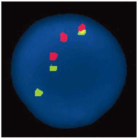
Fig. 3 FISH analysis for an RUNX1/RUNX1T1 probe. Interphase FISH shows an abnormal pattern of three-way variant translocation with two green signals (RUNX1), two red signals (RUNX1T1), and one yellow fusion signal in 98% of the nuclei examined.
Reference
-
1. Cuneo A, Bigoni R, Cavazzini F, Bardi A, Roberti MG, Agostini P, et al. Incidence and significance of cryptic chromosome aberrations detected by fluorescence in situ hybridization in acute myeloid leukemia with normal karyotype. Leukemia. 2002. 16:1745–1751.
Article2. Rowley JD. Identification of a translocation with quinacrine fluorescence in a patient with acute leukemia. Annales Genetiques. 1973. 16:109–112.3. Swerdlow S, Campo E, editors. WHO classification of tumours of haematopoietic and lymphoid tissuses. 2008. 4th ed. Lyon: IARC;1108.4. Kim H, Kim M, Lim J, Kim Y, Han K, Kim SY, et al. A case of acute myeloid leukemia with masked t(8;21). Korean J Lab Med. 2006. 26:338–343.
Article5. Vundinti BR, Kerketta L, Madkaikar M, Jijina F, Ghosh K. Three way translocation in a new variant of t(8;21) acute myeloid leukemia involving Xp22. Indian J Cancer. 2008. 45:30–32.
Article6. Al Bahar S, Adriana Z, Pandita R. A novel variant translocation t(6;8;21)(p22;q22;q22) leading to AML/ETO fusion in acute myeloid leukemia. Gulf J Oncolog. 2009. 5:56–59.7. Miyagi J, Kakazu N, Masuda M, Miyagi T, Toyohama T, Nakazato T, et al. Acute myeloid leukemia (FAB-M2) with a masked type of t(8;21) translocation revealed by spectral karyotyping. Int J Hematol. 2002. 76:338–343.
Article8. Harrison CJ, RadfordWeiss I, Ross F, Rack K, le Guyader G, Vekemans M, et al. Fluorescence in situ hybridization analysis of masked (8;21) (q22;q22) translocations. Cancer Genet Cytogenet. 1999. 112:15–20.
Article9. Kawakami K, Nishii K, Hyou R, Watanabe Y, Nakao M, Mitani H, et al. A case of acute myeloblastic leukemia with a novel variant of t(8;21) (q22;q22). Int J Hematol. 2008. 87:78–82.
Article10. Onozawa M, Fukuhara T, Nigo M, Takeda A, Takahata M, Yamamoto Y, et al. Insertion (21;8)(q22;q22q22): a masked t(8;21) in a patient with acute myelocytic leukemia. Cancer Genet Cytogenet. 2003. 147:134–139.
Article11. Soenen V, Preudhomme C, Roumier C, Daudignon A, Laï JL, Fenaux P. 17p Deletion in acute myeloid leukemia and myelodysplastic syndrome. Analysis of breakpoints and deleted segments by fluorescence in situ. Blood. 1998. 91:1008–1015.
Article12. Im M, Lee JK, Lee DY, Hong YJ, Hong SI, Kang HJ, et al. Near-tetraploidy acute myeloid leukemia with RUNX1-RUNX1T1 rearrangement due to cryptic t(8;21). Korean J Lab Med. 2009. 29:510–514.
Article13. Saitoh K, Miura I, Ohshima A, Takahashi N, Kume M, Utsumi S, et al. Translocation (8;12;21)(q22.1;q24.1;q22.1) a new masked type of t(8;21)(q22;q22) in a patient with acute myeloid leukemia. Cancer Genet Cytogenet. 1997. 96:111–114.
Article14. Udayakumar AM, Alkindi S, Pathare AV, Raeburn HA. Complex t(8;13;21)(q22;q14;q22)--a novel variant of t(8;21) in a patient with acute myeloid leukemia (AML-M2). Arch Med Res. 2008. 39:252–256.
Article15. Vieira L, Oliveira V, Ambrósio AP, Marques B, Pereira AM, Hagemeijer A, et al. Translocation (8;17;15;21)(q22;q23;q15;q22) in acute myeloid leukemia (M2). a fourway variant of t(8;21). Cancer Genet Cytogenetic. 2001. 128:104–107.
- Full Text Links
-
- Actions
-
Cited
- CITED
-
- Close
- Share
-
- Similar articles
-
- Two Concurrent Chromosomal Aberrations Involving Three-way t(3;21;8)(p21;q22;q22) and Two-way t(2;11)(q31;p15) Translocations in a Case of de novo Acute Myeloid Leukemia
- A Case of Acute Myeloid Leukemia with Masked t(8;21)
- A Case of Acute Myeloid Leukemia with an Unusual Phenotype
- Acute Myeloid Leukemia with t(8;21)(q22;q22) (AML1/ETO) in a Patient with Marked Hypocellularity and Low Blasts Count
- The Cytogenetic Analysis of Leukemia Patients; A Study of 515 Cases
