Lab Anim Res.
2013 Sep;29(3):138-147. 10.5625/lar.2013.29.3.138.
Morphometrical dimensions of the sheep thoracolumbar vertebrae as seen on digitised CT images
- Affiliations
-
- 1Large Animal Clinic for Surgery, Faculty of Veterinary Medicine, University of Leipzig, Leipzig, Germany. mahmoud.mageed@hotmail.com
- 2Department of Surgery and Anaesthesia, Faculty of Veterinary Medicine, University of Khartoum, Shambat, Sudan.
- 3Microsurgery and Animal Models Core, Translational Centre for Regenerative Medicine, University of Leipzig, Leipzig, Germany.
- 4Department of Neurosurgery, BG Hospital Bergmannstrost, Halle, Germany.
- KMID: 2312106
- DOI: http://doi.org/10.5625/lar.2013.29.3.138
Abstract
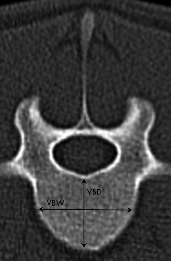
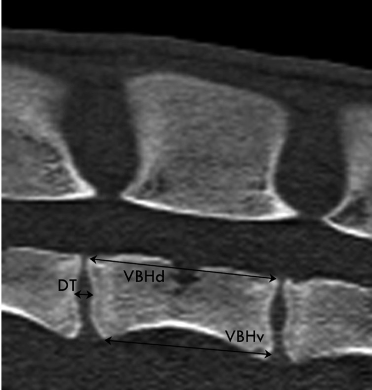
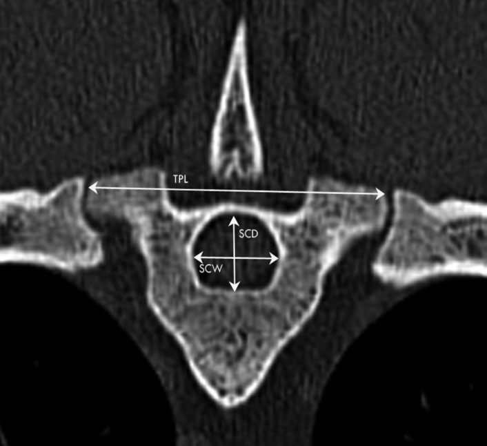
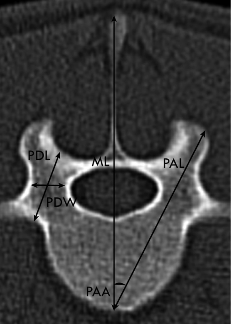
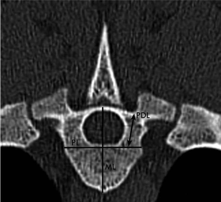
- The sheep spine is widely used as a model for preclinical research in human medicine to test new spinal implants and surgical procedures. Therefore, precise morphometric data are needed. The present study aimed to provide computed tomographic (CT) morphometry of sheep thoracolumbar spine. Five adult normal Merino sheep were included in this study. Sheep were anaesthetised and positioned in sternal recumbency. Subsequently, transverse and sagittal images were obtained using a multi-detector-row helical CT scanner. Measurements of the vertebral bodies, pedicles, intervertebral disc and transverse processes were performed with dedicated software. Vertebral bodies and the spinal canal were wider than they were deep, most obviously in the lumbar vertebrae. The intervertebral discs were as much as 57.4% thicker in the lumbar than in the thoracic spine. The pedicles were higher and longer than they were wide over the entire thoracolumbar spine. In conclusion, the generated data can serve as a CT reference for the ovine thoracolumbar spine and may be helpful in using sheep spine as a model for human spinal research.
MeSH Terms
Figure
Cited by 1 articles
-
Is sheep lumbar spine a suitable alternative model for human spinal researches? Morphometrical comparison study
Mahmoud Mageed, Dagmar Berner, Henriette Jülke, Christian Hohaus, Walter Brehm, Kerstin Gerlach
Lab Anim Res. 2013;29(4):183-189. doi: 10.5625/lar.2013.29.4.183.
Reference
-
1. Wilke HJ, Wenger K, Claes L. Testing criteria for spinal implants: recommendations for the standardization of in vitro stability testing of spinal implants. Eur Spine J. 1998; 7(2):148–154. PMID: 9629939.
Article2. Goel VK, Panjabi MM, Patwardhan AG, Dooris AP, Serhan H. American Society for Testing and Materials. Test protocols for evaluation of spinal implants. J Bone Joint Surg Am. 2006; 88(Suppl 2):103–109. PMID: 16595454.
Article3. Ashman RB, Bechtold JE, Edwards WT, Johnston CE 2nd, McAfee PC, Tencer AF. In vitro spinal arthrodesis implant mechanical testing protocols. J Spinal Disord. 1989; 2(4):274–281. PMID: 2520086.
Article4. Tominaga T, Dickman CA, Sonntag VK, Coons S. Comparative anatomy of the baboon and the human cervical spine. Spine. 1995; 20(2):131–137. PMID: 7716616.
Article5. Smit TH. The use of a quadruped as an in vivo model for the study of the spine - biomechanical considerations. Eur Spine J. 2002; 11(2):137–144. PMID: 11956920.
Article6. Edmondston SJ, Singer KP, Day RE, Breidahl PD, Price RI. Formalin fixation effects on vertebral bone density and failure mechanics: an in-vitro study of human and sheep vertebrae. Clin Biomech (Bristol, Avon). 1994; 9(3):175–179.
Article7. Eggli S, Schläpfer F, Angst M, Witschger P, Aebi M. Biomechanical testing of three newly developed transpedicular multisegmental fixation systems. Eur Spine J. 1992; 1(2):109–116. PMID: 20054957.
Article8. Gurwitz GS, Dawson JM, McNamara MJ, Federspiel CF, Spengler DM. Biomechanical analysis of three surgical approaches for lumbar burst fractures using short-segment instrumentation. Spine. 1993; 18(8):977–982. PMID: 8367785.
Article9. Yoganandan N, Kumaresan S, Voo L, Pintar FA. Finite element applications in human cervical spine modeling. Spine (Phila Pa 1976). 1996; 21(15):1824–1834. PMID: 8855470.
Article10. Kiefer A, Shirazi-Adl A, Parnianpour M. Stability of the human spine in neutral postures. Eur Spine J. 1997; 6(1):45–53. PMID: 9093827.
Article11. Newman E, Turner AS, Wark JD. The potential of sheep for the study of osteopenia: current status and comparison with other animal models. Bone. 1995; 16(4):277S–284S. PMID: 7626315.
Article12. Nunamaker DM. Experimental models of fracture repair. Clin Orthop Relat Res. 1998; 355:S56–S65. PMID: 9917626.
Article13. Bergmann G, Graichen F, Rohlmann A. Hip joint forces in sheep. J Biomech. 1999; 32(8):769–777. PMID: 10433418.
Article14. Egermann M, Goldhahn J, Schneider E. Animal models for fracture treatment in osteoporosis. Osteoporos Int. 2005; 16:S129–S138. PMID: 15750681.
Article15. Turner AS. Experiences with sheep as an animal model for shoulder surgery: strengths and shortcomings. J Shoulder Elbow Surg. 2007; 16(5):S158–S163. PMID: 17507248.
Article16. Krag MH, Weaver DL, Beynnon BD, Haugh LD. Morphometry of the thoracic and lumbar spine related to transpedicular screw placement for surgical spinal fixation. Spine. 1988; 13(1):27–32. PMID: 3381134.
Article17. Olsewski JM, Simmons EH, Kallen FC, Mendel FC, Severin CM, Berens DL. Morphometry of the lumbar spine: anatomical perspectives related to transpedicular fixation. J Bone Joint Surg Am. 1990; 72(4):541–549. PMID: 2139030.18. Abuzayed B, Tutunculer B, Kucukyuruk B, Tuzgen S. Anatomic basis of anterior and posterior instrumentation of the spine: morphometric study. Surg Radiol Anat. 2010; 32(1):75–85. PMID: 19696959.
Article19. Kadioglu HH, Takci E, Levent A, Arik M, Aydin IH. Measurements of the lumbar pedicles in the Eastern Anatolian population. Surg Radiol Anat. 2003; 25(2):120–126. PMID: 12748815.
Article20. Wolf A, Shoham M, Michael S, Moshe R. Morphometric study of the human lumbar spine for operation-workspace specifications. Spine. 2001; 26(22):2472–2477. PMID: 11707713.
Article21. Way TW, Chan HP, Goodsitt MM, Sahiner B, Hadjiiski LM, Zhou C, Chughtai A. Effect of CT scanning parameters on volumetric measurements of pulmonary nodules by 3D active contour segmentation: a phantom study. Phys Med Biol. 2008; 53(5):1295–1312. PMID: 18296763.
Article22. Beers GJ, Carter AP, Leiter BE, Tilak SP, Shah RR. Interobserver discrepancies in distance measurements from lumbar spine CT scans. Am J Roentgenol. 1985; 144(2):395–398. PMID: 3871289.
Article23. Zhou SH, McCarthy ID, McGregor AH, Coombs RR, Hughes SP. Geometrical dimensions of the lower lumbar vertebrae--analysis of data from digitised CT images. Eur Spine J. 2000; 9(3):242–248. PMID: 10905444.24. Martini L, Fini M, Giavaresi G, Giardino R. Sheep model in orthopedic research: a literature review. Comp Med. 2001; 51(4):292–299. PMID: 11924786.25. Kumar N, Kukreti S, Ishaque M, Mulholland R. Anatomy of deer spine and its comparison to the human spine. Anat Rec. 2000; 260(2):189–203. PMID: 10993955.
Article26. McLain RF, Yerby SA, Moseley TA. Comparative morphometry of L4 vertebrae: comparison of large animal models for the human lumbar spine. Spine (Phila Pa 1976). 2002; 27(8):E200–E206. PMID: 11935119.27. Riley LH 3rd, Eck JC, Yoshida H, Koh YD, You JW, Lim TH. A biomechanical comparison of calf versus cadaver lumbar spine models. Spine. 2004; 29(11):E217–E220. PMID: 15167671.
Article28. Wilke HJ, Kettler A, Claes LE. Are sheep spines a valid biomechanical model for human spines? Spine. 1997; 22(20):2365–2374. PMID: 9355217.
Article29. Wilke HJ, Kettler A, Wenger KH, Claes LE. Anatomy of the sheep spine and its comparison to the human spine. Anat Rec. 1997; 247(4):542–555. PMID: 9096794.
Article30. Tins B. Technical aspects of CT imaging of the spine. Insights imaging. 2010; 1(5-6):349–359. PMID: 22347928.
Article31. Mitchell B, Williams J. Respiratory function changes in sheep associated with lying in lateral recumbency and with sedation by xylazine. Vet Anaesth Analg. 1976; 6(1):30–36.
Article32. Schwarz T, Saunders J. CT acquisitation principle. In : Schwarz T, Saunders J, editors. Veterinary computed tomography. 1st ed. Oxford: Wiley-Blackwell;2011. p. 9–27.33. Seiler G, Kinns J, Dennison S, Saunders J, Schwarz T. Vertebral column and spinal cord. In : Schwarz T, Saunders J, editors. Veterinary computed tomography. 1st ed. Oxford: Wiley-Blackwell;2011. p. 209–228.34. Bland JM, Altman DG. Statistical methods for assessing agreement between two methods of clinical measurement. Lancet. 1986; 1(8476):307–310. PMID: 2868172.
Article35. Flynn JR, Bolton PS. Measurement of the vertebral canal dimensions of the neck of the rat with a comparison to the human. Anat Rec. 2007; 290(7):893–899.
Article36. Tatarek NE. Variation in the human cervical neural canal. Spine J. 2005; 5(6):623–631. PMID: 16291101.
Article37. Denoix JM. Spinal biomechanics and functional anatomy. Vet Clin North Am Equine Pract. 1999; 15(1):27–60. PMID: 10218240.
Article38. Schönström N, Lindahl S, Willén J, Hansson T. Dynamic changes in the dimensions of the lumbar spinal canal: an experimental study in vitro. J Orthop Res. 1989; 7(1):115–121. PMID: 2908901.
Article39. Inufusa A, An HS, Lim TH, Hasegawa T, Haughton VM, Nowicki BH. Anatomic changes of the spinal canal and intervertebral foramen associated with flexion-extension movement. Spine. 1996; 21(21):2412–2420. PMID: 8923625.
Article40. Louis R. Fusion of the lumbar and sacral spine by internal fixation with screw plates. Clin Orthop Relat Res. 1986; 203:18–33. PMID: 3955980.
Article41. Roy-Camille R, Saillant G, Mazel C. Internal fixation of the lumbar spine with pedicle screw plating. Clin Orthop Relat Res. 1986; 203:7–17. PMID: 3955999.
Article42. Zindrick MR, Wiltse LL, Doornik A, Widell EH, Knight GW, Patwardhan AG, Thomas JC, Rothman SL, Fields BT. Analysis of the morphometric characteristics of the thoracic and lumbar pedicles. Spine. 1987; 12(2):160–166. PMID: 3589807.
Article
- Full Text Links
- Actions
-
Cited
- CITED
-
- Close
- Share
- Similar articles
-
- Is sheep lumbar spine a suitable alternative model for human spinal researches? Morphometrical comparison study
- Morphologic Feasibility of Pedicle Screw Insertion in Korean
- Lumbar Morphometry: A Study of Lumbar Vertebrae from a Pakistani Population Using Computed Tomography Scans
- Fractal Dimension Of Ct Images Of Normal Parotid Glands
- Measurement of Canal Encroachment Using Axial and Sagittal-Reconstructed Computed Tomographic Images in Thoracolumbar Burst Fractures

