Ann Rehabil Med.
2016 Apr;40(2):334-340. 10.5535/arm.2016.40.2.334.
Visual Evoked Potential Using Head-Mounted Display Versus Cathode Ray Tube: A Pilot Study
- Affiliations
-
- 1Department of Rehabilitation Medicine and Research Institute of Rehabilitation Medicine, Yonsei University College of Medicine, Seoul, Korea. bettertomo@yuhs.ac
- 2Department of Rehabilitation Medicine, CHA Bundang Medical Center, CHA University, Seongnam, Korea.
- 3Department of Physical Medicine and Rehabilitation, Myongji Hospital, Seonam University College of Medicine, Goyang, Korea.
- KMID: 2309934
- DOI: http://doi.org/10.5535/arm.2016.40.2.334
Abstract
OBJECTIVE
To present a new stimulation method based on the use of a head-mounted display (HMD) during pattern reversal visual evoked potential (PR-VEP) testing and to compare variables of HMD to those of conventional cathode ray tube (CRT).
METHODS
Twenty-three normal subjects without visual problems were recruited. PR-VEPs were generated using CRT or HMD stimuli. VEP outcome measures included latencies (N75, P100, and N145) and peak-to-peak amplitudes (N75-P100 and P100-N145). Subjective discomfort associated with HMD was determined using a self-administered questionnaire.
RESULTS
PR-VEPs generated by HMD stimuli showed typical triphasic waveforms, the components of which were found to be correlated with those obtained using conventional CRT stimuli. Self-administered discomfort questionnaires revealed that HMD was more comfortable in some aspects. It allowed subjects to concentrate better than CRT.
CONCLUSION
The described HMD stimulation can be used as an alternative to the standard CRT stimulation for PR-VEPs. PR-VEP testing using HMD has potential applications in clinical practice and visual system research because HMD can be used on a wider range of subjects compared to CRT.
MeSH Terms
Figure
-
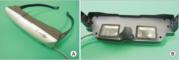
Fig. 1 A picture of head-mounted display. (A) The front side of the unit. (B) An inside view showing the organic light emitting diode (OLED) monitors.
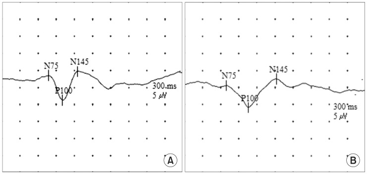
Fig. 2 Waveform of pattern obtained during reversal-visual evoked potential (PR-VEP) testing using. (A) A cathode ray tube (CRT). (B) A head-mounted display (HMD). The PR-VEPs obtained in both CRT and HMD showed well-defined N75, P100, and N145.
Reference
-
1. Cobb WA, Dawson GD. The latency and form in man of the occipital potentials evoked by bright flashes. J Physiol. 1960; 152:108–121. PMID: 13810780.
Article2. Vaughan HG Jr, Hull RC. Functional relation between stimulus intensity and photically evoked cerebral responses in man. Nature. 1965; 206:720–722. PMID: 5832861.
Article3. Odom JV, Bach M, Barber C, Brigell M, Marmor MF, Tormene AP, et al. Visual evoked potentials standard (2004). Doc Ophthalmol. 2004; 108:115–123. PMID: 15455794.
Article4. Jeffrey LC, Goldberg G. Central nervous system electrophysiology. In : DeLisa JA, Gans BM, Walsh NE, editors. Physical medicine and rehabilitation: principles and practice. 4th ed. Philadelphia: Lippincott Williams & Wilkins;2005. p. 105–137.5. Velger M. Helmet-mounted displays and sights. Boston: Artech House;1998. p. 10–15.6. Hanprasertpong C, Koizumi Y, Aoyagi M, Kimura M, Yagi T. A head-mounted visual stimulator for neurotological examination. Auris Nasus Larynx. 2004; 31:379–382. PMID: 15571910.
Article7. Melzer JE, Moffitt K. HMD design: putting the user first. In : Melzer JE, Moffitt K, editors. Head mounted displays: designing for the user. New York: McGraw-Hill;1997. p. 1–16.8. International Organization for Standardization. International standard-ergonomic requirements for office work with visual display terminals (VDTs). Part 3: visual display requirements. Geneva: International Organization for Standardization;1992. (ISO 9241-3:1992).9. Oosterhuis HJ, Ponsen L, Jonkman EJ, Magnus O. The average visual response in patients with cerebrovascular disease. Electroencephalogr Clin Neurophysiol. 1969; 27:23–34. PMID: 4182886.
Article10. Klassen AC, Heaney LM, Lee MC, Torres F. Hypercapnic alteration of visual evoked responses in acute cerebral infarction. Arch Neurol. 1979; 36:627–629. PMID: 485892.
Article11. Misulis KE, Fakhoury T. Spehlmann's evoked potential primer. 3rd ed. Boston: Butterworth-Heinemann;2001. p. 137–155.12. Bodis-Wollner I, Ghilardi MF, Mylin LH. The importance of stimulus selection in VEP practice: the clinical relevance of visual physiology. In : Cracco RQ, Bodis-Wollner , editors. Evoked potentials. New York: Liss;1986. p. 15–27.13. Celesia GG, DeMarco PJ Jr. Anatomy and physiology of the visual system. J Clin Neurophysiol. 1994; 11:482–492. PMID: 7844239.
Article14. Brigell M, Bach M, Barber C, Kawasaki K, Kooijman A. Guidelines for calibration of stimulus and recording parameters used in clinical electrophysiology of vision. Calibration Standard Committee of the International Society for Clinical Electrophysiology of Vision (ISCEV). Doc Ophthalmol. 1998; 95:1–14. PMID: 10189178.15. Celesia GG. Evoked potential techniques in the evaluation of visual function. J Clin Neurophysiol. 1984; 1:55–76. PMID: 6400020.
Article16. Tobimatsu S, Celesia G, Cone S. Effects of pupil diameter and luminance changes on pattern electroretinograms and visual evoked potentials. Clin Vision Sci. 1988; 2:293–302.17. Tobimatsu S, Kurita-Tashima S, Nakayama-Hiromatsu M, Akazawa K, Kato M. Age-related changes in pattern visual evoked potentials: differential effects of luminance, contrast and check size. Electroencephalogr Clin Neurophysiol. 1993; 88:12–19. PMID: 7681386.
Article18. Kurita-Tashima S, Tobimatsu S, Nakayama-Hiromatsu M, Kato M. Effect of check size on the pattern reversal visual evoked potential. Electroencephalogr Clin Neurophysiol. 1991; 80:161–166. PMID: 1713147.
Article19. Patterson R, Winterbottom MD, Pierce BJ. Perceptual issues in the use of head-mounted visual displays. Hum Factors. 2006; 48:555–573. PMID: 17063969.
Article
- Full Text Links
-
- Actions
-
Cited
- CITED
-
- Close
- Share
-
- Similar articles
-
- The Effect of Using Head Mounted Display on Human Eyes
- Study for Analysis of the Multifocal Visual Evoked Potential
- HVL Measurement of the Miniature X-Ray Tube Using Diode Detector
- Usefulness of Computerized Objective Visual Acuity Test System Using Suppression Method
- Visual Evoked Potentials for Detecting Visual Pathway Abnormality
