Korean J Ophthalmol.
2011 Oct;25(5):334-340. 10.3341/kjo.2011.25.5.334.
Study for Analysis of the Multifocal Visual Evoked Potential
- Affiliations
-
- 1Department of Ophthalmology, Soonchunhyang University Bucheon Hospital, Soonchunhyang University College of Medicine, Bucheon, Korea. yhohn@schbc.ac.kr
- 2Department of Ophthalmology, Soonchunhyang University Gumi Hospital, Soonchunhyang University College of Medicine, Gumi, Korea.
- KMID: 1098636
- DOI: http://doi.org/10.3341/kjo.2011.25.5.334
Abstract
- PURPOSE
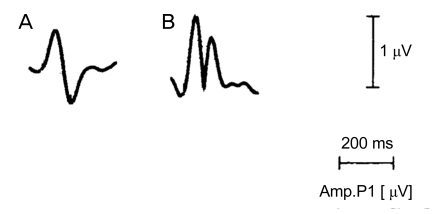
To introduce the clinical utility of the absolute value of the reconstructed waveform method in the analysis of multifocal visual evoked potential (mfVEP).
METHODS
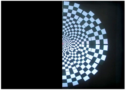
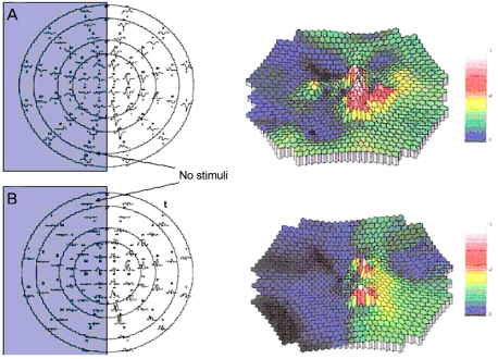
The mfVEP with 4-channel recording was performed using RETIscan(R) on 10 eyes of 10 normal subjects. Amplitudes were obtained from ring-shaped 6 areas and 4 sectors. The best visual evoked potential (VEP) response method and the absolute value of the reconstructed waveform method were compared in terms of analysis of the amplitudes. In order to assess the false positive rate of the examination, stimuli were administered with one-half of the cathode ray tube (CRT) monitor completely covered and the results were compared using 2 methods.
RESULTS
The amplitudes in 6 areas and 4 sectors analyzed with the best VEP response method and the absolute value of the reconstructed waveform method showed no statistical difference (p > 0.05). The amplitude in the stimuli-blocked area of the absolute value of the reconstructed waveform method was smaller than that of the best VEP response method (p < 0.05) and the amplitude of the stimuli area showed no substantial difference between two methods (p > 0.05).
CONCLUSIONS
The absolute value of the reconstructed waveform method has similar reproducibility and lower level of false positives relative to the best VEP response method. Therefore, it can be considered as a useful method in the analysis of the mfVEP.
Keyword
MeSH Terms
Figure
Reference
-
1. Sutter EE. Imaging visual function with the multifocal m-sequence technique. Vision Res. 2001; 41:1241–1255. PMID: 11322969.
Article2. Hood DC, Zhang X, Greenstein VC, et al. An interocular comparison of the multifocal VEP: a possible technique for detecting local damage to the optic nerve. Invest Ophthalmol Vis Sci. 2000; 41:1580–1587. PMID: 10798679.3. Graham SL, Klistorner AI, Grigg JR, Billson FA. Objective VEP perimetry in glaucoma: asymmetry analysis to identify early deficits. J Glaucoma. 2000; 9:10–19. PMID: 10708226.
Article4. Hood DC, Odel JG, Zhang X. Tracking the recovery of local optic nerve function after optic neuritis: a multifocal VEP study. Invest Ophthalmol Vis Sci. 2000; 41:4032–4038. PMID: 11053309.5. Kardon R, Givre S, Wall M, Hood D. Wall M, Mills RP, editors. Comparison of threshold and multifocal VEP perimetry in recovered optic neuritis. Perimetry update 2000/2001. 2001. Amsterdam: Kugler;p. 19–28.6. Hood DC, Odel JG, Winn BJ. The multifocal visual evoked potential. J Neuroophthalmol. 2003; 23:279–289. PMID: 14663311.
Article7. Klistorner A, Graham SL. Objective perimetry in glaucoma. Ophthalmology. 2000; 107:2283–2299. PMID: 11097611.8. Odom JV, Bach M, Brigell M, et al. ISCEV standard for clinical visual evoked potentials (2009 update). Doc Ophthalmol. 2010; 120:111–119. PMID: 19826847.
Article9. Halliday AM, McDonald WI, Mushin J. Delayed visual evoked response in optic neuritis. Lancet. 1972; 1:982–985. PMID: 4112367.
Article10. Halliday AM, McDonald WI, Mushin J. Visual evoked response in diagnosis of multiple sclerosis. Br Med J. 1973; 4:661–664. PMID: 4758547.
Article11. Halliday AM, Michael WF. Changes in pattern-evoked responses in man associated with the vertical and horizontal meridians of the visual field. J Physiol. 1970; 208:499–513. PMID: 5533451.
Article12. Michael WF, Halliday AM. Differences between the occipital distribution of upper and lower field pattern-evoked responses in man. Brain Res. 1971; 32:311–324. PMID: 5134583.
Article13. Ossenblok P, Spekreijse H. The extrastriate generators of the EP to checkerboard onset. A source localization approach. Electroencephalogr Clin Neurophysiol. 1991; 80:181–193. PMID: 1713149.
Article14. Fortune B, Hood DC. Conventional pattern-reversal VEPs are not equivalent to summed multifocal VEPs. Invest Ophthalmol Vis Sci. 2003; 44:1364–1375. PMID: 12601070.
Article15. Howe JW, Mitchell KW. Visual evoked cortical potential to paracentral retinal stimulation in chronic glaucoma, ocular hypertension, and an age-matched group of normals. Doc Ophthalmol. 1986; 63:37–44. PMID: 3732011.
Article16. Hood DC, Greenstein VC, Odel JG, et al. Visual field defects and multifocal visual evoked potentials: evidence of a linear relationship. Arch Ophthalmol. 2002; 120:1672–1681. PMID: 12470141.17. Hood DC, Zhang X, Hong JE, Chen CS. Quantifying the benefits of additional channels of multifocal VEP recording. Doc Ophthalmol. 2002; 104:303–320. PMID: 12076018.18. Slotnick SD, Klein SA, Carney T, et al. Using multi-stimulus VEP source localization to obtain a retinotopic map of human primary visual cortex. Clin Neurophysiol. 1999; 110:1793–1800. PMID: 10574294.
Article19. Hood DC, Zhang X, Winn BJ. Detecting glaucomatous damage with multifocal visual evoked potentials: how can a monocular test work? J Glaucoma. 2003; 12:3–15. PMID: 12567104.
Article20. Fortune B, Zhang X, Hood DC, et al. Normative ranges and specificity of the multifocal VEP. Doc Ophthalmol. 2004; 109:87–100. PMID: 15675203.
Article21. Zhang X, Hood DC. A principal component analysis of multifocal pattern reversal VEP. J Vis. 2004; 4:32–43. PMID: 14995897.
Article22. Chen JY, Hood DC, Odel JG, Behrens MM. The effects of retinal abnormalities on the multifocal visual evoked potential. Invest Ophthalmol Vis Sci. 2006; 47:4378–4385. PMID: 17003429.
Article23. Zhang X, Hood DC, Chen CS, Hong JE. A signal-to-noise analysis of multifocal VEP responses: an objective definition for poor records. Doc Ophthalmol. 2002; 104:287–302. PMID: 12076017.
- Full Text Links
- Actions
-
Cited
- CITED
-
- Close
- Share
- Similar articles
-
- The Change of Multifocal Visual Evoked Potential in Unilateral Anisometropic Amblyopia before and after Occlusion Treatment
- Multifocal Visual Evoked Potential in Normal Subjects
- Visual Evoked Potentials for Detecting Visual Pathway Abnormality
- The Effects of Intravenous Anesthetics on Visual Evoked Potentials in Cats
- Visual evoked potential study in normal neonates

