J Breast Cancer.
2016 Jun;19(2):210-213. 10.4048/jbc.2016.19.2.210.
An Innovative Ultrasound Technique for Evaluation of Tumor Vascularity in Breast Cancers: Superb Micro-Vascular Imaging
- Affiliations
-
- 1Department of Radiology, Korea University Ansan Hospital, Korea University College of Medicine, Ansan, Korea. seoboky@korea.ac.kr
- KMID: 2308974
- DOI: http://doi.org/10.4048/jbc.2016.19.2.210
Abstract
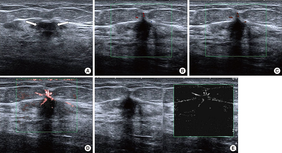
- Tumor vascularity is an important indicator for differential diagnosis, tumor growth, and prognosis. Superb micro-vascular imaging (SMI) is an innovative ultrasound technique for vascular examination that uses a multidimensional filter to eliminate clutter and preserve extremely low-velocity flows. Theoretically, SMI could depict more vessels and more detailed vascular morphology, due to the increased sensitivity of slow blood flow. Here, we report the early experience of using SMI in 21 breast cancer patients. We evaluated tumor vascular features in breast cancer and compared SMI and conventional color or power Doppler imaging. SMI was superior to color or power Doppler imaging in detecting tumor vessels, the details of vessel morphology, and both peripheral and central vascular distribution. In conclusion, SMI is a promising ultrasound technique for evaluating microvascular information of breast cancers.
Keyword
Figure
Cited by 1 articles
-
A Prospective Study on the Value of Ultrasound Microflow Assessment to Distinguish Malignant from Benign Solid Breast Masses: Association between Ultrasound Parameters and Histologic Microvessel Densities
Ah Young Park, Myoungae Kwon, Ok Hee Woo, Kyu Ran Cho, Eun Kyung Park, Sang Hoon Cha, Sung Eun Song, Ju-Han Lee, JaeHyung Cha, Gil Soo Son, Bo Kyoung Seo
Korean J Radiol. 2019;20(5):759-772. doi: 10.3348/kjr.2018.0515.
Reference
-
1. Drudi FM, Cantisani V, Gnecchi M, Malpassini F, Di Leo N, de Felice C. Contrast-enhanced ultrasound examination of the breast: a literature review. Ultraschall Med. 2012; 33:E1–E7.
Article2. Huber S, Helbich T, Kettenbach J, Dock W, Zuna I, Delorme S. Effects of a microbubble contrast agent on breast tumors. Computer-assisted quantitative assessment with color Doppler US: early experience. Radiology. 1998; 208:485–489.
Article3. Giuseppetti GM, Baldassarre S, Marconi E. Color Doppler sonography. Eur J Radiol. 1998; 27:Suppl 2. S254–S258.
Article4. Kook SH, Park HW, Lee YR, Lee YU, Pae WK, Park YL. Evaluation of solid breast lesions with power Doppler sonography. J Clin Ultrasound. 1999; 27:231–237.
Article5. Schroeder RJ, Bostanjoglo M, Rademaker J, Maeurer J, Felix R. Role of power Doppler techniques and ultrasound contrast enhancement in the differential diagnosis of focal breast lesions. Eur Radiol. 2003; 13:68–79.
Article6. Superb micro-vascular imaging (SMI). Toshiba Medical System. Accessed August 18th, 2015. https://medical.toshiba.com/products/ultrasound/aplio-platinum/technology.php.7. Mendelson EB, Böhm-Vélez M, Berg WA, Whitman GJ, Feldman MI, Madjar H, et al. ACR BI-RADS ultrasound. In : D'Orsi CJ, Sickles EA, Mendelson EB, Morris EA, editors. ACR-BI-RADS Atlas: Breast Imaging Reporting and Data System. 5th ed. Reston: American College of Radiology;2013.8. Lee SW, Choi HY, Baek SY, Lim SM. Role of color and power Doppler imaging in differentiating between malignant and benign solid breast masses. J Clin Ultrasound. 2002; 30:459–464.
Article9. Sorelli PG, Cosgrove DO, Svensson WE, Zaman N, Satchithananda K, Barrett NK, et al. Can contrast-enhanced sonography distinguish benign from malignant breast masses? J Clin Ultrasound. 2010; 38:177–181.
Article10. Du J, Wang L, Wan CF, Hua J, Fang H, Chen J, et al. Differentiating benign from malignant solid breast lesions: combined utility of conventional ultrasound and contrast-enhanced ultrasound in comparison with magnetic resonance imaging. Eur J Radiol. 2012; 81:3890–3899.
Article11. Wu L, Yen HH, Soon MS. Spoke-wheel sign of focal nodular hyperplasia revealed by superb micro-vascular ultrasound imaging. QJM. 2015; 108:669–670.
Article12. Ma Y, Li G, Li J, Ren WD. The diagnostic value of superb microvascular imaging (SMI) in detecting blood flow signals of breast lesions: a preliminary study comparing SMI to color Doppler flow imaging. Medicine (Baltimore). 2015; 94:e1502.13. Lee WJ, Chu JS, Huang CS, Chang MF, Chang KJ, Chen KM. Breast cancer vascularity: color Doppler sonography and histopathology study. Breast Cancer Res Treat. 1996; 37:291–298.
Article
- Full Text Links
- Actions
-
Cited
- CITED
-
- Close
- Share
- Similar articles
-
- Up-to-date Doppler techniques for breast tumor vascularity: superb microvascular imaging and contrast-enhanced ultrasound
- A Technical Note on a Novel Technique for the Evaluation of Breast Excision Specimen: Automated Breast Ultrasound System
- Superb microvascular imaging technology of ultrasound examinations for the evaluation of tumor vascularity in hepatic hemangiomas
- Optical Imaging of the Breast
- Real-Time MRI Navigated Ultrasound for Preoperative Tumor Evaluation in Breast Cancer Patients: Technique and Clinical Implementation


