Imaging Sci Dent.
2016 Jun;46(2):109-115. 10.5624/isd.2016.46.2.109.
Prevalence of bony septa, antral pathology, and dimensions of the maxillary sinus from a sinus augmentation perspective: A retrospective cone-beam computed tomography study
- Affiliations
-
- 1Department of Oral and Maxillofacial Radiology, University of Connecticut, School of Dental Medicine, Farmington, CT, USA. tadinada@uchc.edu
- 2Section of Periodontology, University of Connecticut, School of Dental Medicine, Farmington, CT, USA.
- 3Department of Prosthodontics, University of Connecticut, School of Dental Medicine, Farmington, CT, USA.
- 4Department of Pediatric Dentistry, University of Connecticut, School of Dental Medicine, Farmington, CT, USA.
- 5Division of Periodontology, University of Dammam, College of Dentistry, Dammam, Saudi Arabia.
- KMID: 2308874
- DOI: http://doi.org/10.5624/isd.2016.46.2.109
Abstract
- PURPOSE
Sinus elevation procedures have become a routine and reliable way to gain bone volume in the edentulous maxilla for dental implant placement. Presence of bony septations and pathology in the maxillary sinus often cause complications leading to graft or implant failure or both. The aim of this study was to retrospectively evaluate the prevalence of pathology, direction of the septa, and sinus width measured at 2 mm, 5 mm, and 10 mm from the sinus floor in maxillary sinuses using cone-beam computed tomography (CBCT).
MATERIALS AND METHODS
Seventy-two sinuses from 36 random preoperative CBCT scans referred for implant therapy were retrospectively evaluated for the number, prevalence, and direction of bony septations and presence of pathology. Width of the sinus was also measured at 2 mm, 5 mm, and 10 mm from the sinus floor to account for the amount of bone available for implant placement.
RESULTS
Maxillary sinus septa were found in 59.7%. Presence of a single septum was noted in 20 sinuses (27.7%), followed by two septa in 17 sinuses. The most common direction of the septum was the transverse direction. Retention pseudocyst and mucosal thickening were the most commonly seen abnormality/pathology.
CONCLUSION
Based on the high prevalence of septa and sinus pathology in this sample, a preoperative CBCT scan might be helpful in minimizing complications during sinus augmentation procedures for dental implant therapy.
MeSH Terms
Figure
-
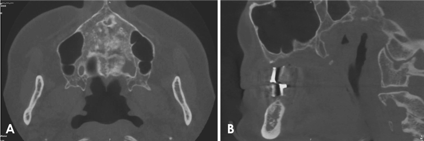
Fig. 1 Septa are seen on an axial (A) and coronal (B) section images.
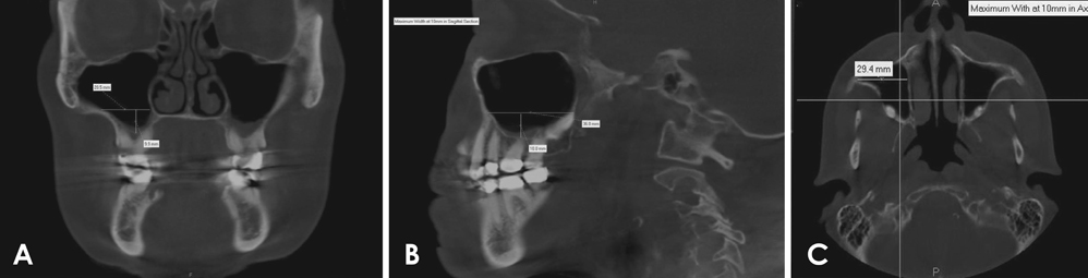
Fig. 2 A. Maximum disto-medial dimension is measured at 10 mm cranial to the floor of the sinus in coronal section. B. Maximum antero-posterior dimensions at 10 mm cranial to the floor of the sinus is measured on the sagittal section. C. Maximum width at 10 mm cranial to the floor of the sinus is measured in axial section.
Reference
-
1. Tadinada A, Fung K, Thacker S, Mahdian M, Jadhav A, Schincaglia GP. Radiographic evaluation of the maxillary sinus prior to dental implant therapy: a comparison between two-dimensional and three-dimensional radiographic imaging. Imaging Sci Dent. 2015; 45:169–174.
Article2. Balshi TJ, Wolfinger GJ. Management of the posterior maxilla in the compromised patient: historical, current, and future perspectives. Periodontol 2000. 2003; 33:67–81.
Article3. Chappard D, Baslé MF, Legrand E, Audran M. Trabecular bone microarchitecture: a review. Morphologie. 2008; 92:162–170.
Article4. Sharan A, Madjar D. Maxillary sinus pneumatization following extractions: a radiographic study. Int J Oral Maxillofac Implants. 2008; 23:48–56.5. Raghoebar GM, Timmenga NM, Reintsema H, Stegenga B, Vissink A. Maxillary bone grafting for the insertion of endosseous implants: results after 12-124 months. Clin Oral Implants Res. 2001; 12:279–286.6. Peleg M, Garg AK, Mazor Z. Predictability of simultaneous implant placement in the severely atrophic posterior maxilla: a 9-year longitudinal experience study of 2132 implants placed into 731 human sinus grafts. Int J Oral Maxillofac Implants. 2006; 21:94–102.
Article7. Nedir R, Nurdin N, Vazquez L, Szmukler-Moncler S, Bischof M, Bernard JP. Osteotome sinus floor elevation technique without grafting: a 5-year prospective study. J Clin Periodontol. 2010; 37:1023–1028.
Article8. Blus C, Szmukler-Moncler S, Salama M, Salama H, Garber D. Sinus bone grafting procedures using ultrasonic bone surgery: 5-year experience. Int J Periodontics Restorative Dent. 2008; 28:221–229.9. Pjetursson BE, Tan WC, Zwahlen M, Lang NP. A systematic review of the success of sinus floor elevation and survival of implants inserted in combination with sinus floor elevation. J Clin Periodontol. 2008; 35:8 Suppl. 216–240.
Article10. Bornstein MM, Chappuis V, von Arx T, Buser D. Performance of dental implants after staged sinus floor elevation procedures: 5-year results of a prospective study in partially edentulous patients. Clin Oral Implants Res. 2008; 19:1034–1043.
Article11. Wallace SS, Froum SJ. Effect of maxillary sinus augmentation on the survival of endosseous dental implants. A systematic review. Ann Periodontol. 2003; 8:328–343.
Article12. Emmerich D, Att W, Stappert C. Sinus floor elevation using osteotomes: a systematic review and meta-analysis. J Periodontol. 2005; 76:1237–1251.
Article13. Galindo-Moreno P, Avila G, Fernández-Barbero JE, Aguilar M, Sánchez-Fernández E, Cutando A, et al. Evaluation of sinus floor elevation using a composite bone graft mixture. Clin Oral Implants Res. 2007; 18:376–382.
Article14. Marino MJ, Weinstein JE, Riley CA, Levy JM, Emerson NA, McCoul ED. Assessment of pneumatization of the paranasal sinuses: a comprehensive and validated metric. Int Forum Allergy Rhinol. 2016; 6:429–436.
Article15. Yoo JY, Pi SH, Kim YS, Jeong SN, You HK. Healing pattern of the mucous membrane after tooth extraction in the maxillary sinus. J Periodontal Implant Sci. 2011; 41:23–29.
Article16. Tiwana PS, Kushner GM, Haug RH. Maxillary sinus augmentation. Dent Clin North Am. 2006; 50:409–424.
Article17. Testori T, Weinstein RL, Taschieri S, Del Fabbro M. Risk factor analysis following maxillary sinus augmentation: a retrospective multicenter study. Int J Oral Maxillofac Implants. 2012; 27:1170–1176.18. Kennedy DW, Zinreich SJ, Rosenbaum AE, Johns ME. Functional endoscopic sinus surgery. Theory and diagnostic evaluation. Arch Otolaryngol. 1985; 111:576–582.
Article19. Mafee MF, Chow JM, Meyers R. Functional endoscopic sinus surgery: anatomy, CT screening, indications, and complications. AJR Am J Roentgenol. 1993; 160:735–744.
Article20. Maestre-Ferrín L, Galán-Gil S, Rubio-Serrano M, Peñarrocha-Diago M, Peñarrocha-Oltra D. Maxillary sinus septa: a systematic review. Med Oral Patol Oral Cir Bucal. 2010; 15:e383–e386.21. Wen SC, Chan HL, Wang HL. Classification and management of antral septa for maxillary sinus augmentation. Int J Periodontics Restorative Dent. 2013; 33:509–517.
Article22. Călin C, Petre A, Drafta S. Osteotome-mediated sinus floor elevation: a systematic review and meta-analysis. Int J Oral Maxillofac Implants. 2014; 29:558–576.
Article23. Benavides E, Rios HF, Ganz SD, An CH, Resnik R, Reardon GT, et al. Use of cone beam computed tomography in implant dentistry: the International Congress of Oral Implantologists consensus report. Implant Dent. 2012; 21:78–86.24. Schwartz-Arad D, Herzberg R, Dolev E. The prevalence of surgical complications of the sinus graft procedure and their impact on implant survival. J Periodontol. 2004; 75:511–516.
Article25. Barone A, Santini S, Sbordone L, Crespi R, Covani U. A clinical study of the outcomes and complications associated with maxillary sinus augmentation. Int J Oral Maxillofac Implants. 2006; 21:81–85.26. Ziccardi VB, Betts NJ. Complications of maxillary sinus augmentation. In : Jensen OT, editor. The sinus bone graft. Chicago, IL: Quintessence;1999. p. 201–208.27. Regev E, Smith RA, Perrott DH, Pogrel MA. Maxillary sinus complications related to endosseous implants. Int J Oral Maxillofac Implants. 1995; 10:451–461.28. Gosau M, Rink D, Driemel O, Draenert FG. Maxillary sinus anatomy: a cadaveric study with clinical implications. Anat Rec (Hoboken). 2009; 292:352–354.
Article29. Ulm CW, Solar P, Krennmair G, Matejka M, Watzek G. Incidence and suggested surgical management of septa in sinus-lift procedures. Int J Oral Maxillofac Implants. 1995; 10:462–465.30. Velásquez-Plata D, Hovey LR, Peach CC, Alder ME. Maxillary sinus septa: a 3-dimensional computerized tomographic scan analysis. Int J Oral Maxillofac Implants. 2002; 17:854–860.31. González-Santana H, Peñarrocha-Diago M, Guarinos-Carbó J, Sorní-Bröker M. A study of the septa in the maxillary sinuses and the subantral alveolar processes in 30 patients. J Oral Implantol. 2007; 33:340–343.
Article32. Krennmair G, Ulm C, Lugmayr H. Maxillary sinus septa: incidence, morphology and clinical implications. J Craniomaxillofac Surg. 1997; 25:261–265.
Article
- Full Text Links
-
- Actions
-
Cited
- CITED
-
- Close
- Share
-
- Similar articles
-
- Maxillary sinus septa: comparison between panoramic radiography and CBCT
- Comparison of panoramic radiography and cone beam computed tomography for assessing the relationship between the maxillary sinus floor and maxillary molars
- Prevalence of incidental paranasal sinus opacification in dental paediatric patients
- Maxillary sinus aspergilloma of odontogenic origin: Report of 2 cases with cone-beam computed tomographic findings and review of the literature
- Assessment of the relationship between the maxillary molars and adjacent structures using cone beam computed tomography
