Pregnancy in Hypertrophic Cardiomyopathy with Severe Left Ventricular Outflow Tract Obstruction
- Affiliations
-
- 1Department of Cardiovascular Medicine, Gachon University Gil Medical Center, Incheon, Korea. heart@gachon.ac.kr
- 2Gachon Cardiovascular Research Institute, Gachon University, Incheon, Korea.
- KMID: 2308706
- DOI: http://doi.org/10.4250/jcu.2016.24.2.158
Abstract
- Hypertrophic obstructive cardiomyopathy (HOCM) patients with severe left ventricular outflow tract (LVOT) obstruction (those with a gradient of > 100 mm Hg) are at the highest risk of hemodynamic deterioration during pregnancy. Complications of HOCM include sudden cardiac death, heart failure, and arrhythmias. Physiological changes during pregnancy may induce these complications, affecting maternal and fetal health conditions. Therefore, close monitoring with appropriate management is essential for the well-being of both mother and fetus. We report on the case of a 27-year-old female patient with severe LVOT obstruction HOCM, pressure gradient (PG) of 125 mm Hg at resting, and 152 mm Hg induced by the Valsalva maneuver at 34 weeks gestation. This case showed how close monitoring using echocardiography and proper management during the course of pregnancy resulted in successful delivery in the patient with extremely high PG HOCM.
Keyword
MeSH Terms
Figure
-
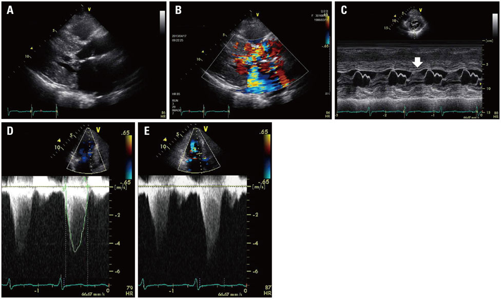
Fig. 1 Baseline transthoracic echocardiography (TTE). TTE showed massive asymmetric septal hypertrophy of left ventricular septum with wall thickness > 22 mm in parasternal long axis view (A), eccentric mitral regurgitation grade III/IV (B), systolic anterior motion (white arrow) of mitral valve presented by M-mode (C), very high left ventricular outflow tract pressure gradient at resting (D), and during the Valsalva maneuver (E).
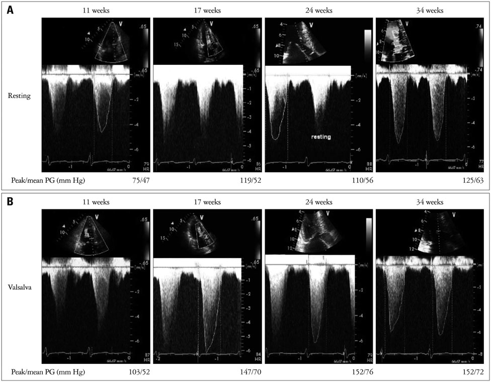
Fig. 2 Serial check up of left ventricular outflow tract (LVOT) pressure gradient (PG) presented by M-mode at resting (A) and during the Valsalva maneuver (B). At 11 weeks gestation, initial transthoracic echocardiography examination showed LVOT PG (peak/mean PG; 75/47 mm Hg at rest, 103/52 mm Hg during Valsalva maneuver). As the pregnancy progresses, when 34 weeks gestation, LVOT PG was increasing up to 125/63 mm Hg at resting and 152/72 mm Hg on the Valsalva maneuver.
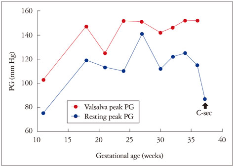
Fig. 3 Serial follow up of left ventricular outflow tract (LVOT) pressure during pregnancy. This graph shows the serial values of LVOT PG at resting (blue line) and during the Valsalva maneuver (red line) which changes according to the gestational age. PG: pressure gradient.
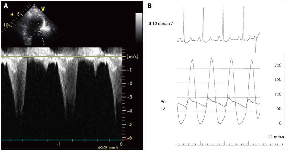
Fig. 4 Transthoracic echocardiography (TTE) and cardiac catheterization just before the alcohol septal ablation. TTE showed severe left ventricular outflow tract (LVOT) pressure gradient (PG) (A) and cardiac catheterization showed also high LVOT PG (127 mm Hg) at the same time (B). Ao: aorta, LV: left ventricle.
Reference
-
1. Maron BJ. Hypertrophic cardiomyopathy: a systematic review. JAMA. 2002; 287:1308–1320.2. Tanaka H, Kamiya C, Katsuragi S, Tanaka K, Miyoshi T, Tsuritani M, Yoshida M, Iwanaga N, Neki R, Yoshimatsu J, Ikeda T. Cardiovascular events in pregnancy with hypertrophic cardiomyopathy. Circ J. 2014; 78:2501–2506.3. Shim WJ. Role of echocardiography in the management of cardiac disease in women. J Cardiovasc Ultrasound. 2014; 22:173–179.4. Alegria JR, Nishimura R. Hypertrophic cardiomyopathy and pregnancy. In : Oakley C, Warnes CA, editors. Heart disease in pregnancy. Oxford: Blackwell Publishing;2007. p. 173–185.5. Maron BJ, Gardin JM, Flack JM, Gidding SS, Kurosaki TT, Bild DE. Prevalence of hypertrophic cardiomyopathy in a general population of young adults. Echocardiographic analysis of 4111 subjects in the CARDIA Study. Coronary Artery Risk Development in (Young) Adults. Circulation. 1995; 92:785–789.6. Thaman R, Varnava A, Hamid MS, Firoozi S, Sachdev B, Condon M, Gimeno JR, Murphy R, Elliott PM, McKenna WJ. Pregnancy related complications in women with hypertrophic cardiomyopathy. Heart. 2003; 89:752–756.7. Pieper PG, Walker F. Pregnancy in women with hypertrophic cardiomyopathy. Neth Heart J. 2013; 21:14–18.8. European Society of Gynecology (ESG). Association for European Paediatric Cardiology (AEPC). German Society for Gender Medicine (DGesGM). Regitz-Zagrosek V, Blomstrom Lundqvist C, Borghi C, Cifkova R, Ferreira R, Foidart JM, Gibbs JS, Gohlke-Baerwolf C, Gorenek B, Iung B, Kirby M, Maas AH, Morais J, Nihoyannopoulos P, Pieper PG, Presbitero P, Roos-Hesselink JW, Schaufelberger M, Seeland U, Torracca L. ESC Committee for Practice Guidelines. ESC Guidelines on the management of cardiovascular diseases during pregnancy: the Task Force on the Management of Cardiovascular Diseases during Pregnancy of the European Society of Cardiology (ESC). Eur Heart J. 2011; 32:3147–3197.9. Krul SP, van der Smagt JJ, van den Berg MP, Sollie KM, Pieper PG, van Spaendonck-Zwarts KY. Systematic review of pregnancy in women with inherited cardiomyopathies. Eur J Heart Fail. 2011; 13:584–594.10. Gersh BJ, Maron BJ, Bonow RO, Dearani JA, Fifer MA, Link MS, Naidu SS, Nishimura RA, Ommen SR, Rakowski H, Seidman CE, Towbin JA, Udelson JE, Yancy CW. American College of Cardiology Foundation/American Heart Association Task Force on Practice Guidelines. 2011 ACCF/AHA Guideline for the Diagnosis and Treatment of Hypertrophic Cardiomyopathy: a report of the American College of Cardiology Foundation/American Heart Association Task Force on Practice Guidelines. Developed in collaboration with the American Association for Thoracic Surgery, American Society of Echocardiography, American Society of Nuclear Cardiology, Heart Failure Society of America, Heart Rhythm Society, Society for Cardiovascular Angiography and Interventions, and Society of Thoracic Surgeons. J Am Coll Cardiol. 2011; 58:e212–e260.
- Full Text Links
-
- Actions
-
Cited
- CITED
-
- Close
- Share
-
- Similar articles
-
- Biventricular Hypertrophic Cardiomyopathy with Severe Right Ventricular Outflow Track Obstruction
- Hypertrophic cardiomyopathy secondary to severe right and left ventricular outflow tract obstruction in a Maltese dog
- A Case of Normalized Hypertrophic Cardiomyopathy after Removal of Pheochromocytoma
- Flail Subaortic Membrane Mimicking Left Ventricular Outflow Tract Obstruction in Hypertrophic Cardiomyopathy
- The Change of Left Ventricular outflow Tract before and after Myomymectomy of Hypertrophic Obstructive Cardiomyopathy
