J Cardiovasc Ultrasound.
2013 Jun;21(2):90-93.
Flail Subaortic Membrane Mimicking Left Ventricular Outflow Tract Obstruction in Hypertrophic Cardiomyopathy
- Affiliations
-
- 1Regional Cardiovascular Center, Department of Internal Medicine, Chungnam National University Hospital, Chungnam National University School of Medicine, Daejeon, Korea. jojeong@cnu.ac.kr
Abstract
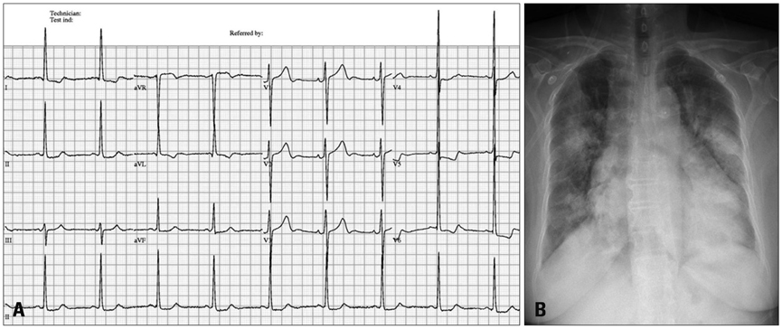
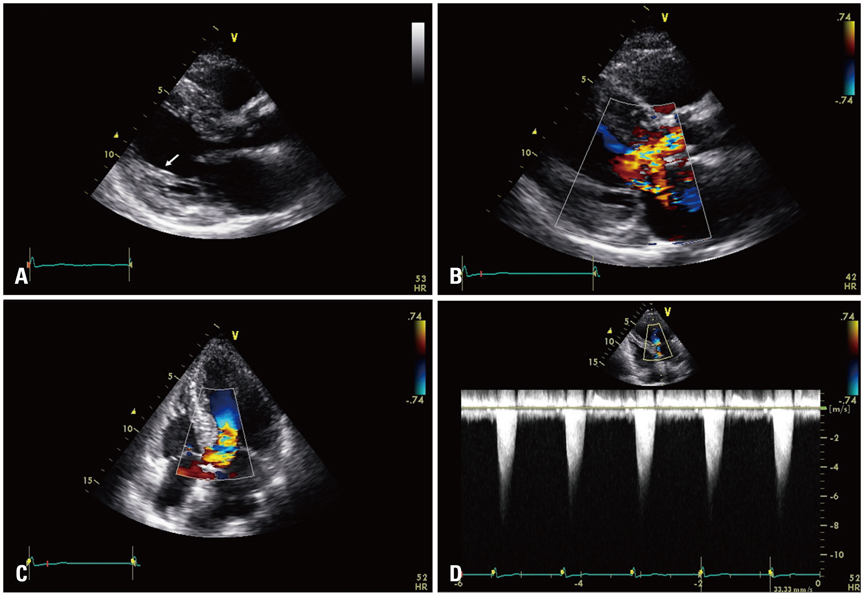
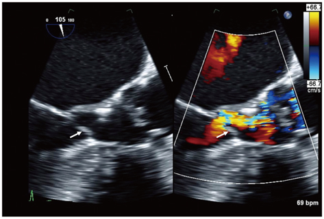
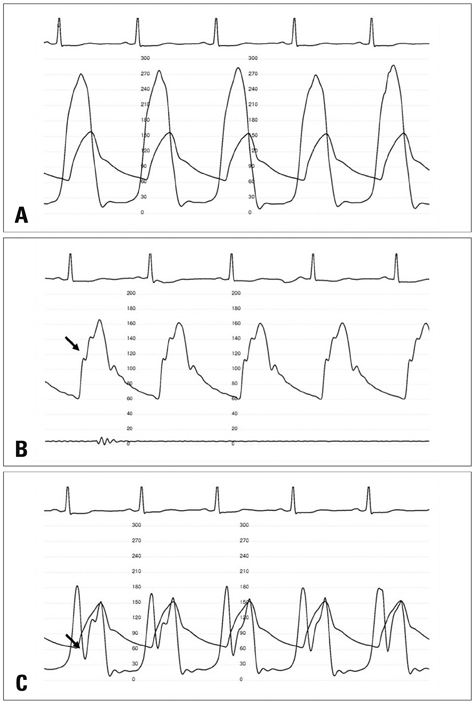
- A subaortic membrane is an uncommon cause for left ventricular outflow tract obstruction. Hypertrophic cardiomyopathy with dynamic left ventricular outflow tract obstruction would mask the presence of the subaortic membrane on transthoracic echocardiography and cause a false diagnosis. We report a patient with subaortic stenosis due to flail subaortic membrane misdiagnosed as obstructive hypertrophic cardiomyopathy on transthoracic echocardiography, identified on transesophageal echocardiography and cardiac catheterization.
MeSH Terms
Figure
Reference
-
1. Firpo C, Maitre Azcárate MJ, Quero Jiménez M, Saravalli O. Discrete subaortic stenosis (DSS) in childhood: a congenital or acquired disease? Follow-up in 65 patients. Eur Heart J. 1990; 11:1033–1040.
Article2. Choi JY, Sullivan ID. Fixed subaortic stenosis: anatomical spectrum and nature of progression. Br Heart J. 1991; 65:280–286.
Article3. Teis A, Sheppard MN, Alpendurada F. Subaortic membrane: correlation of imaging with pathology. Eur Heart J. 2010; 31:2822.
Article4. Maron MS, Olivotto I, Betocchi S, Casey SA, Lesser JR, Losi MA, Cecchi F, Maron BJ. Effect of left ventricular outflow tract obstruction on clinical outcome in hypertrophic cardiomyopathy. N Engl J Med. 2003; 348:295–303.
Article5. Bruce CJ, Nishimura RA, Tajik AJ, Schaff HV, Danielson GK. Fixed left ventricular outflow tract obstruction in presumed hypertrophic obstructive cardiomyopathy: implications for therapy. Ann Thorac Surg. 1999; 68:100–104.
Article6. Oliver JM, González A, Gallego P, Sánchez-Recalde A, Benito F, Mesa JM. Discrete subaortic stenosis in adults: increased prevalence and slow rate of progression of the obstruction and aortic regurgitation. J Am Coll Cardiol. 2001; 38:835–842.
Article7. Onbasili AO, Tekten T, Ceyhan C. Subaortic stenosis caused by flail discrete membrane in an older patient. Heart. 2004; 90:399.
Article8. Nagueh SF, Bierig SM, Budoff MJ, Desai M, Dilsizian V, Eidem B, Goldstein SA, Hung J, Maron MS, Ommen SR, Woo A. American Society of Echocardiography. American Society of Nuclear Cardiology. Society for Cardiovascular Magnetic Resonance. Society of Cardiovascular Computed Tomography. American Society of Echocardiography clinical recommendations for multimodality cardiovascular imaging of patients with hypertrophic cardiomyopathy: Endorsed by the American Society of Nuclear Cardiology, Society for Cardiovascular Magnetic Resonance, and Society of Cardiovascular Computed Tomography. J Am Soc Echocardiogr. 2011; 24:473–498.
Article
- Full Text Links
- Actions
-
Cited
- CITED
-
- Close
- Share
- Similar articles
-
- Biventricular Hypertrophic Cardiomyopathy with Severe Right Ventricular Outflow Track Obstruction
- Multiplane Transesophageal Echocardiographic Findings of Two Cases of Discrete Subvalvular Aortic Stenosis
- A Case of Normalized Hypertrophic Cardiomyopathy after Removal of Pheochromocytoma
- Epidural Anesthesia in Patient with Idiopathic Hypertrophic Subaortic Stenosis
- Hypertrophic cardiomyopathy secondary to severe right and left ventricular outflow tract obstruction in a Maltese dog


