Korean J Leg Med.
2013 Aug;37(3):134-138. 10.7580/kjlm.2013.37.3.134.
A Study on Mineral Changes on the Weathering Human Hair after Burial using EDX
- Affiliations
-
- 1Department of Anatomy and Cell Biology, College of Medicine, Hanyang University, Seoul, Korea.
- 2Department of Biomedical Engineering, College of Health Science, Eulji University, Seongnam-si, Gyeonggi, Korea.
- 3Department of Anatomy, Catholic Institute for Applied Anatomy, College of Medicine, Catholic University, Seoul, Korea.
- 4Division of Forensic Medicine, National Forensic Service, Seoul, Korea.
- 5Department of Funeral Science, College of Health Industry, Eulji University, Seongnam-si, Gyeonggi, Korea. hks@eulji.ac.kr
- KMID: 2305465
- DOI: http://doi.org/10.7580/kjlm.2013.37.3.134
Abstract
- This study was undertaken to investigate mineral changes in weathered scalp hair after burial. EDX (energy dispersive X-ray spectroscopy) analysis was performed to measure the presence of minerals on the hair surface. Twelve scalp hairs, buried for 5-40 years, were chosen from deceased individuals buried in tombs in Soha-Ri, Kyonggi-Do, and other regions in Korea. Three normal hairs were used as the control group. EDX data showed that carbon, oxygen, and sulfur were detected in hair collected from all three burial grounds. In contrast, calcium was only detected in hair collected from tombs in Soha-ri. The amounts of calcium and sulfur were found to decrease with time for hair collected from tombs in Soha-ri. Similar results were observed with sodium for hair collected from other regions. These results show region specific mineral detection and a decrease in the concentration of minerals with time. Consequently, it is suggested that changes in minerals concentration in weathered hair could be used as basic data in the field of forensic medicine.
Keyword
MeSH Terms
Figure
-
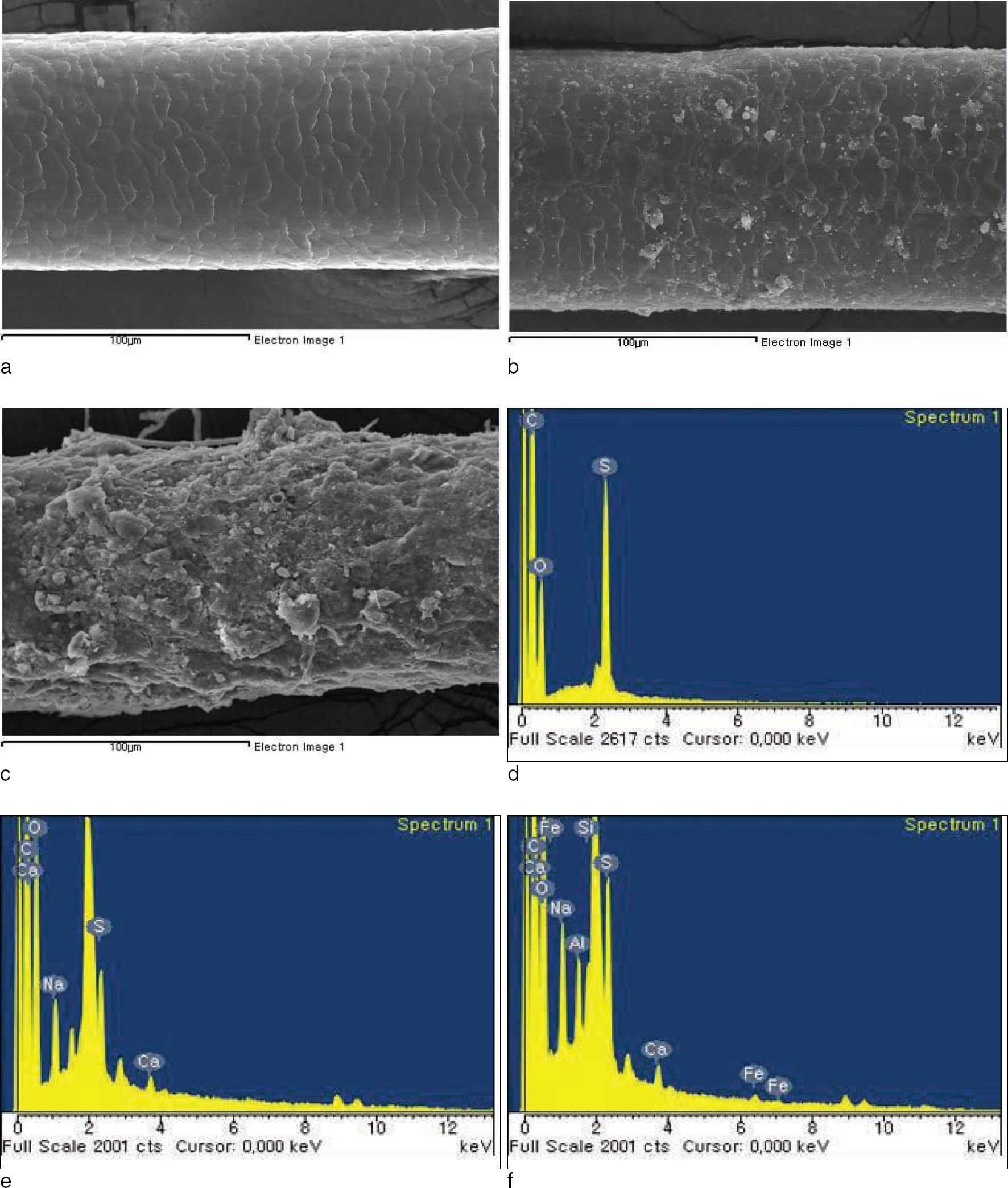
Fig. 1. SEM images of the surface and typical EDX analysis spectra are shown. The morphological changes in weathering hair shafts are investigated by SEM (control hair (a), ≤ 10 years after burial (b) and ≥30 years after burial (c)). EDX to analyze the material found in the surface of the control hair (d), ≤ 10 years after burial (e) and ≥30 years after burial (f). C (Carbon), O (Oxygen), S (Sulfur), Na (Sodium), Ca (Calcium), Al (Aluminium) and Fe (iron).
Reference
-
1. Amory S, Keyser-Tracqui C, Crubezy E, et al. Multi-sub-strata analysis on Siberian mummies: a different way for validation in ancient DNA Studies? Int Congr Ser. 2006; 1288:834–6.
Article2. Daniel CR 3rd, Piraccini BM, Tosti A. The nail and hair in forensic science. J Am Acad Dermatol. 2004; 50:258–61.
Article3. Wilson AS, Dixon RA, Dodson HI, et al. Yesterday’ s hair-human hair in archaeology. Biologist (London). 2001; 48:213–7.4. Chang BS, Uhm CS, Park CH, et al. Ultramicroscopic study on the hair of newly found 15th century mummy in Daejeon, Korea. Ann Anat. 2006; 188:439–45.
Article5. Hwang KS, Nam YS, Kim JL, et al. TEM Observation on the weathering human scalp hair after burial. Korean J Phys Anthropol. 2012; 25:1–10.
Article6. Hwang KS, Lim DS, Ahn DC, et al. SEM observation on the weathering human hair after burial. Korean J Phys Anthropol. 2008; 21:181–90.
Article7. Ratnapandian S, Warner SB, Kamath YK. Photodegradation of human hair. J Cosmet Sci. 1998; 49:309–20.8. Kempson IM, Skinner WM, Kirkbride PK, et al. Time-of-flight secondary ion mass spectrometry analysis of hair from archaeological remains. Eur J Mass Spectrom. 2003; 9:589–97.
Article9. Lee GY, Chang BS. Studies on the preservative condition and the ultrastructure of hair of newly found sixteenth century mummy in Paju. Korean J Electron Microscopy. 2005; 35:211–8.10. Kempson IM, Skinner WM, Kirkbride PK. Calcium distributions in human hair by ToF-SIMS. Biochim Biophys Acta. 2003; 1624:1–5.
Article11. Dawber R, Comaish S. Scanning electron microscopy of normal and abnormal hair shafts. Arch Dermatol. 1970; 101:315–22.
Article12. Giehl KA, Ferguson DJ, Dawber RP, et al. Update on detection, morphology and fragility in pili annulati in three kindreds. J Eur Acad Dermatol Venereol. 2004; 18:654–8.
Article13. Venning VA, Dawber RP, Ferguson DJ, et al. Weathering of hair in trichothiodystrophy. Br J Dermatol. 1986; 114:591–5.
Article14. Whiting DA. Structural abnormalities of the hair shaft. J Am Acad Dermatol. 1987; 16:1–25.
Article14. Rook A. The clinical importance of ‘weathering’ in human hair. Br J Dermatol. 1976; 95:111–2.
Article16. Scanavez C, Silveria M, Joekes I. Human hair: color changes caused by daily care damage on ultra-structure. Colloids Surf B Biointerfaces. 2003; 28:39–52.17. Du AY, Mangelson NF, Rees LB, et al. PIXE elemental analysis of South American mummy hair. Nucl Instrum Methods Phys Res B. 1996; 109/110:673–6.
Article18. Moretto P, Surleve-Bazeille JE, Licu D, et al. Microanalysis of the human skin structure: preliminary results. Nucl Instrum Methods Phys Res B. 1999; 158:386–92.
Article
- Full Text Links
-
- Actions
-
Cited
- CITED
-
- Close
- Share
-
- Similar articles
-
- SEM Observation on the Weathering Human Hair after Burial
- TEM Observation on the Weathering Human Scalp Hair after Burial
- Four Cases of Hair Shaft Breakage Caused by Hair Care Cosmetics
- Reliability on Intra-Laboratory and Inter-Laboratory Data of Hair Mineral Analysis Comparing with Blood Analysis
- Human Skeletal Remains from Ancient Burial Sites in India: With Special Reference to Harappan Civilization