Korean J Sports Med.
2012 Jun;30(1):34-40. 10.5763/kjsm.2012.30.1.34.
Surgical Excision of Symptomatic Non United Fragment of Anterior Process Fractures of the Calcaneus
- Affiliations
-
- 1Korea National Sport University, Seoul, Korea.
- 2Surgery of Foot and Ankle, Eulji General Hospital, Eulji University, College of Medicine, Seoul, Korea. jins33@hanmail.net
- 3KT Lee's Foot and Ankle Clinic, Seoul, Korea.
- KMID: 2288646
- DOI: http://doi.org/10.5763/kjsm.2012.30.1.34
Abstract
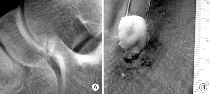
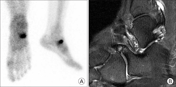
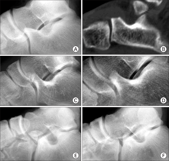
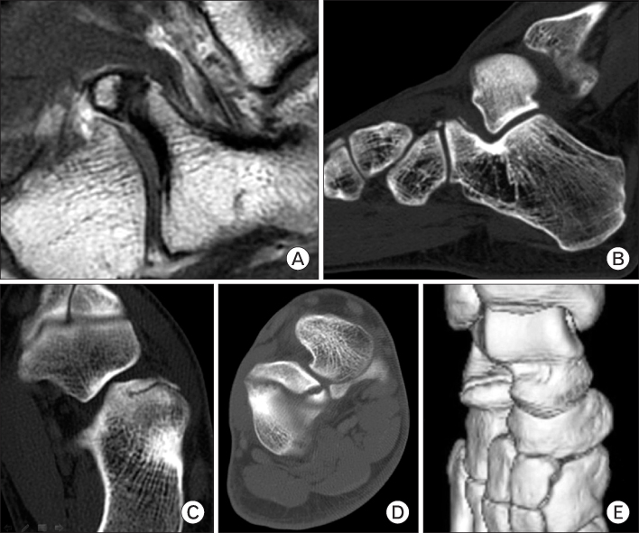
- A fracture of the anterior process of the calcaneus has been considered unusual injury. A clinically missed diagnosis is often, that had gone on to non united fragment. Particularly if the patient has calcaneocuboid pain and disability, and that early excision of the fragment is usually advisable. There were 12 cases with performing the simple excision. The fracture characteristics were analyzed by Degan's classification; type 1 was 1case (8.3%), type 2 was 6 cases (50.0%) and type 3 was 5 cases (41.7%); and their morphology; elongation was 3 cases (50.0%) and fragmentation 3 cases (50.0%). And, the pre and post operative American Orthopedic Foot and Ankle Society midfoot score and visual analog scale was evaluated; 66.0 and 5.8 was significantly improved to 90.1 (p=0.007) and 2.2 (p=0.003), respectively. Postoperative Excellent and good satisfaction with possible return to previous sports activity was 10 cases (83.3%). Conclusively, simple excision of non united fragment of anterior process of the calcaneus is a successful clinical option.
Figure
Reference
-
1. Morel J. Varietes Anatomo-clinique et Prognostic des Fractures du Calcaneum. 1904. Lyon:2. Jahss MH, Kay BS. An anatomic study of the anterior superior process of the os calcis and its clinical application. Foot Ankle. 1983. 3:268–281.3. Petrover D, Schweitzer ME, Laredo JD. Anterior process calcaneal fractures: a systematic evaluation of associated conditions. Skeletal Radiol. 2007. 36:627–632.4. Warrick CK, Bremner AE. Fractures of the calcaneum, with an atlas illustrating the various types of fracture. J Bone Joint Surg Br. 1953. 35:33–45.5. Duddy RK, Donahue WE Jr, Cavolo DJ. Anterior calcaneal process fractures. Recognition and treatment. J Am Podiatry Assoc. 1984. 74:398–401.6. Ouellette H, Salamipour H, Thomas BJ, Kassarjian A, Torriani M. Incidence and MR imaging features of fractures of the anterior process of calcaneus in a consecutive patient population with ankle and foot symptoms. Skeletal Radiol. 2006. 35:833–837.7. Degan TJ, Morrey BF, Braun DP. Surgical excision for anterior-process fractures of the calcaneus. J Bone Joint Surg Am. 1982. 64:519–524.8. Trnka HJ, Zettl R, Ritschl P. Fracture of the anterior superior process of the calcaneus: an often misdiagnosed fracture. Arch Orthop Trauma Surg. 1998. 117:300–302.9. Agnholt J, Nielsen S, Christensen H. Lesion of the ligamentum bifurcatum in ankle sprain. Arch Orthop Trauma Surg. 1988. 107:326–328.10. Kitaoka HB, Alexander IJ, Adelaar RS, Nunley JA, Myerson MS, Sanders M. Clinical rating systems for the ankle-hindfoot, midfoot, hallux, and lesser toes. Foot Ankle Int. 1994. 15:349–353.11. Wulker N, Zwipp H, Tscherne H. Experimental study of the classification of intra-articular calcaneus fractures. Unfallchirurg. 1991. 94:198–203.12. Golder WA. Anterior process of the calcaneus: a clinical-radiological contribution to anatomical vocabulary. Surg Radiol Anat. 2004. 26:163–166.13. Robbins MI, Wilson MG, Sella EJ. MR imaging of anterosuperior calcaneal process fractures. AJR Am J Roentgenol. 1999. 172:475–479.14. Backman S, Johnson SR. Torsion of the foot causing fracture of the anterior calcaneal process. Acta Chir Scand. 1953. 105:460–466.15. Pouliquen JC, Duranthon LD, Glorion C, Kassis B, Langlais J. The too-long anterior process calcaneus: a report of 39 cases in 25 children and adolescents. J Pediatr Orthop. 1998. 18:333–336.16. Pearce CJ, Zaw H, Calder JD. Stress fracture of the anterior process of the calcaneus associated with a calcaneonavicular coalition: a case report. Foot Ankle Int. 2011. 32:85–88.17. Hodge JC. Anterior process fracture or calcaneus secundarius: a case report. J Emerg Med. 1999. 17:305–309.18. Bradford CH, Larsen I. Sprain-fractures of the anterior lip of the os calcis. N Engl J Med. 1951. 244:970–972.
- Full Text Links
- Actions
-
Cited
- CITED
-
- Close
- Share
- Similar articles
-
- Surgical Treatment for Displaced Intra-Articular Calcaneal Fractures
- Penetration of Joints by Screws on Anterior Process of Calcaenus
- Intra-Articular Fractures of the Calcaneus: Open reduction and internal fixation via extended lateral transcalcaneal approach
- Arthroscopically assisted Minimal Invasive Surgery of Calcaneal Intraarticular Fractures
- Treatment of Secondary Soft Tissue Compromised Calcaneus Fractures Using a Cannulated Screw and Simple Cerclage Wiring: A Report of Two Cases


