J Breast Cancer.
2007 Dec;10(4):278-281. 10.4048/jbc.2007.10.4.278.
Localized Polyarteritis Nodosa of the Breast with Mammary Duct Ectasia: A Case Report
- Affiliations
-
- 1Department of Pathology, University of Ulsan College of Medicine, Asan Medical Center, Seoul, Korea.
- 2Department of Pathology, University of Ulsan College of Medicine, Ulsan University Hospital, Ulsan, Korea. heej0124@medimail.co.kr
- 3Department of Radiology, University of Ulsan College of Medicine, Ulsan University Hospital, Ulsan, Korea.
- KMID: 2286598
- DOI: http://doi.org/10.4048/jbc.2007.10.4.278
Abstract
- We describe here a case of localized polyarteritis nodosa that involved the unilateral breast in a 69-yr-old woman. She presented with a tender breast mass and had suffered for two months. On physical examination, an ill-defined 2 cm sized, firm mass was palpated. Ultrasonographic examination revealed a mass like lesion that contained microcalcifications. The mass was excised because of the suspicion of carcinoma. The histologic findings were vasculitis involving medium and small sized-arteries that showed marked neutrophilic and lymphocytic infiltrations with intimal fibroplasias and fragmentation of the internal elastic lamina. The patient progressed well after surgical excision. The discussion includes the importance of differential diagnosis between localized polyarteritis nodosa and other vasculitis, and review of previously reported cases of vasculitis of the breast. Only 13 cases of polyarteritis nodosa of the breast have been reported and this is the first case of polyarteritis nodosa with mammary duct ectasia.
Keyword
MeSH Terms
Figure
-

Fig 1 Ultrasonogram: An ill-defined heterogenous and irregular shaped mass-like lesion is present. Multifocal microcalcification is apparent (arrows).

Fig 2 Small to medium sized-artery show vasculitis in a background of ductal ectasia (Hematoxilin-Eosin stain, ×100).
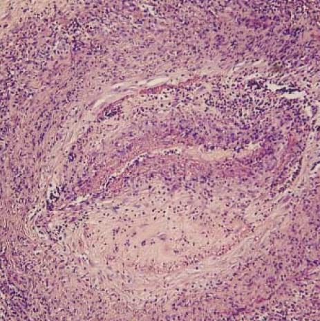
Fig 3 The blood vessel wall is partly obliterated by the inflammatory cells with fibrin thrombi (Hematoxilin-Eosin stain, ×400).
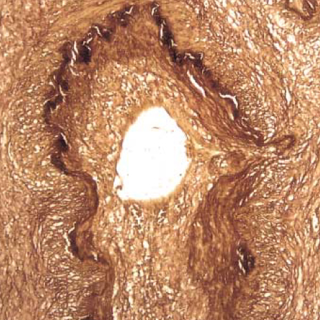
Fig 4 Marked necrotizing vasculitis with fragmentation of internal elastic lamina and calcification are identified in the vessel wall (Elastic stain, ×400).
Reference
-
1. Trueb RM, Scheidegger EPS, Percin M, Singh A, Hoffmann U, Burg G, et al. Periarteritis nodosa presenting as a breast lesion: report of a case and review of the literature. Br J Dermatol. 1999. 141:1117–1121.
Article2. Dega FJ, Hunder GG. Vasculitis of the breast: an unusual manifestation of polyarteritis. Arthritis Rheum. 1974. 17:973–976.
Article3. Park CH, Kang K, Pai ST. Polyarteritis nodosa of the breast. J Korean Soc Surg. 1991. 41:544–548.4. Kim DM, Kim SK, Chang MC, Myong NH, Herr H, Chang HK. A limited polyarteritis nodosa of the breast. J Korean Rheum Assoc. 2005. 12:57–60.5. Ng WF, Chow LT, Lam PW. Localized polyarteritis nodosa of breast-report of two cases and a review of the literature. Histopathology. 1993. 23:535–539.
Article6. Kariv R, Sidi Y, Gur H. Systemic vasculitis presenting as a tumor like lesion: four case reports and an analysis of 79 reported cases. Medicine (Baltimore). 2000. 79:349–359.
Article7. Niles JL. Value of tests for antineutrophil cytoplasmic autoantibodies in the diagnosis and treatment of vasculitis. Curr Opin Rheumatol. 1993. 5:18–24.
Article8. Bajema IM, Hagen EC. Evolving concepts about the role of antineutrophil cytoplasmic autoantibodies in systemic vasculitis. Curr Opin Rheumatol. 1999. 11:34–40.
Article9. Carson CW, Conn DL, Czaja AJ, Wright TL, Brecher ME. Frequency and significance of antibodies to hepatitis C virus in polyarteritis nodosa. J Rheumatol. 1993. 20:304–309.10. Leurez-Ville M, Lauge A, Morinet F, Guillevin L, Deny P. Polyarteritis nodosa and parvovirus B19. Lancet. 1994. 344:263–264.11. Sergent JS, Lockshin MD, Christian CL, Gocke DJ. Vasculitis with hepatitis B antigenemia: Long-term observations in nine patients. Medicine (Baltimore). 1976. 55:1–18.12. Thomas RH, Black MM. The wide clinical spectrum of polyarteritis nodosa with cutaneous involvement. Clin Exp Dermatol. 1983. 8:47–59.
Article13. Bohrod MG, Bodon GF. Isolated polyarteritis nodosa of the gallbladder. Am Surg. 1970. 36:681–685.14. Ansell ID, Evans DJ, Wright DGD. Asymptomatic arteritis of the uterine cervix. J Clin Path. 1974. 27:664–668.
Article15. McCarty DJ, Imbrigia J, Hung JK. Vasculitis of the breast. Arthritis Rheum. 1968. 11:796–803.16. Yamashina M, Wilson TK. A mammographic finding in focal polyarteritis nodosa. Br J Radiol. 1985. 58:91–92.
Article17. PetersEn L, Graversen HP, Andersen JA, Dyreborg U, Blichert-Toft M. The duct ectasia syndrome-an overlooked disease entity. Ugeskr Laeger. 1993. 155:1540–1545.18. Sasmano A, Roseman D, Haber MH. Giant cell arteritis of the breast. A unique syndrome. Arch Intern Med. 1990. 150:900–904.
Article19. Trub RM, Pericin M, Kohler E, Barandum J, Burg G. Necrotizing granulomatosis of the breast. Br J Dermatol. 1997. 137:799–803.
Article20. Johnson WC, Wallrich R, Helwig EB. Superficial thrombophlebitis of the chest wall: Mondor's disease. JAMA. 1962. 180:103–108.
- Full Text Links
-
- Actions
-
Cited
- CITED
-
- Close
- Share
-
- Similar articles
-
- A Benign Biliary Stricture Complicating Isolated Polyarteritis Nodosa
- Polyarteritis nodosa of the breast
- Polyarteritis Nodosa Localized in Small Intestine: A Case Report
- Ischemic Pseudomembranous Colitis with Perforation due to Polyarteritis Nodosa
- Mammary duct ectasia with bloody nipple discharge in a child
