J Bone Metab.
2014 Aug;21(3):205-212. 10.11005/jbm.2014.21.3.205.
Efficacy of Dual Energy X-ray Absorptiometry for Evaluation of Biomechanical Properties: Bone Mineral Density and Actual Bone Strength
- Affiliations
-
- 1Department of Medical and Health Science, Gyeongju University, Gyeongju, Korea.
- 2Department of Biomedical Sciences, Kyungpook National University, Daegu, Korea.
- 3Department of Orthopaedic Surgery, Kyungpook National University School of Medicine, Daegu, Korea. ihpark@knu.ac.kr
- KMID: 2286290
- DOI: http://doi.org/10.11005/jbm.2014.21.3.205
Abstract
- INTRODUCTION
Bone mineral density (BMD) is an important index in diagnosis of osteoporosis and other metabolic bone diseases, prediction of fractures, and monitoring treatment. This study was to find a more feasible technique for prediction of osteoporotic fracture between dual energy X-ray absorptiometry (DXA) and quantitative computed tomography (QCT) and to reveal the actual change of bone strength when BMD was changed.
METHODS
Ten of these 20 specimens were used as the demineralized group and the other 10 as the control. Each specimen was immersed in HCl solution at for a period of at least 10 minutes, up to 100 minutes, at an interval of 10 minutes for different levels of demineralization. BMD was measured using DXA and QCT. Uniaxial compression tests were conducted to measure biomechanical parameters. Pearson correlation analysis was used respectively between BMD and biomechanical parameters and between DXA and QCT.
RESULTS
Elastic modulus (r=0.87) and yield stress (r=0.84) showed a statistically significant correlation with DXA BMD. Through correlation analysis with QCT BMD and elastic modulus, correlation coefficient showed hemi-vertebra (r=0.80) and trabecular (r=0.68). In yield stress, there was a statistically significant correlation in hemi-vertebra (r=0.87) and trabecular bone (r=0.84).
CONCLUSION
DXA is a current standard technique not only for diagnosis of osteoporosis but also for prediction of fracture risk compared to QCT. Actual decrease of bone strength was much greater than that of BMD by both DXA and QCT.
MeSH Terms
Figure
-

Fig. 1 (A) All specimens were cut off (approximately 15×10×15 mm3) in the shape of a cube and (B) were compressed under one-loaded platen rods on an axial compression testing machine in displacement control.
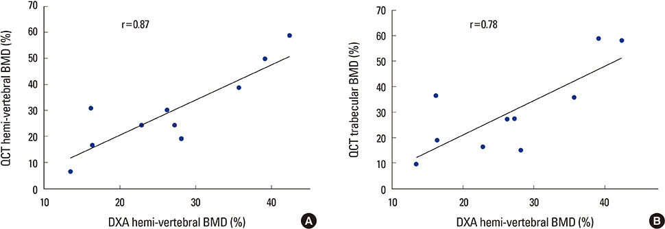
Fig. 2 The graphs showed a strong correlation (A) between quantitative computed tomography (QCT) hemi-vertebral bone mineral density (BMD) and dual energy X-ray absorptiometry (DXA) BMD (r=0.87), (B) QCT trabecular and DXA BMD (r=0.78) as time progression.
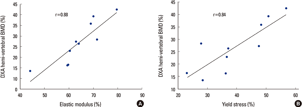
Fig. 3 The graphs showed a strong correlation (A) between elastic modulus (r=0.88) for dual energy X-ray absorptiometry (DXA) bone mineral density (BMD) and (B) yield stress and quantitative computed tomography (QCT) hemi-vertebral BMD (r=0.87) as time progression.
Reference
-
1. Consensus development conference: diagnosis, prophylaxis, and treatment of osteoporosis. Am J Med. 1993; 94:646–650.2. Ettinger B, Black DM, Nevitt MC, et al. The Study of Osteoporotic Fractures Research Group. Contribution of vertebral deformities to chronic back pain and disability. J Bone Miner Res. 1992; 7:449–456.
Article3. Lee YK, Jang S, Jang S, et al. Mortality after vertebral fracture in Korea: analysis of the National Claim Registry. Osteoporos Int. 2012; 23:1859–1865.4. Damilakis J, Maris TG, Karantanas AH. An update on the assessment of osteoporosis using radiologic techniques. Eur Radiol. 2007; 17:1591–1602.
Article5. Liu S, Oguchi Y, Borner JW, et al. Increased canine pancreatic acinar cell damage after organophosphate and acetylcholine or cholecystokinin. Pancreas. 1990; 5:177–182.
Article6. Gregg EW, Kriska AM, Salamone LM, et al. The epidemiology of quantitative ultrasound: a review of the relationships with bone mass, osteoporosis and fracture risk. Osteoporos Int. 1997; 7:89–99.
Article7. Bergot C, Laval-Jeantet AM, Hutchinson K, et al. A comparison of spinal quantitative computed tomography with dual energy X-ray absorptiometry in European women with vertebral and nonvertebral fractures. Calcif Tissue Int. 2001; 68:74–82.
Article8. Black DM, Bouxsein ML, Marshall LM, et al. Proximal femoral structure and the prediction of hip fracture in men: a large prospective study using QCT. J Bone Miner Res. 2008; 23:1326–1333.
Article9. Engelke K, Adams JE, Armbrecht G, et al. Clinical use of quantitative computed tomography and peripheral quantitative computed tomography in the management of osteoporosis in adults: the 2007 ISCD Official Positions. J Clin Densitom. 2008; 11:123–162.
Article10. Lee CY, Chan SH, Lai HY, et al. A method to develop an in vitro osteoporosis model of porcine vertebrae: histological and biomechanical study. J Neurosurg Spine. 2011; 14:789–798.
Article11. Actis AB, Obwegeser JA, Rupérez C. Influence of different sterilization procedures and partial demineralization of screws made of bone on their mechanical properties. J Biomater Appl. 2004; 18:193–207.
Article12. McLean FC, Urist MR. Bone, an introduction to the physiology of skeletal tissue. Chicago, IL: The University of Chicago Press;1955.13. Bayraktar HH, Morgan EF, Niebur GL, et al. Comparison of the elastic and yield properties of human femoral trabecular and cortical bone tissue. J Biomech. 2004; 37:27–35.
Article14. Nelson HD, Helfand M, Woolf SH, et al. Screening for postmenopausal osteoporosis: a review of the evidence for the U.S. Preventive Services Task Force. Ann Intern Med. 2002; 137:529–541.
Article15. Schneider P, Börner W. Indication for bone density measurement and critical review. Nuklearmediziner. 1993; 13:83–91.16. Block JE, Smith R, Glueer CC, et al. Models of spinal trabecular bone loss as determined by quantitative computed tomography. J Bone Miner Res. 1989; 4:249–257.
Article17. Goldstein SA. The mechanical properties of trabecular bone: dependence on anatomic location and function. J Biomech. 1987; 20:1055–1061.
Article18. Goulet RW, Goldstein SA, Ciarelli MJ, et al. The relationship between the structural and orthogonal compressive properties of trabecular bone. J Biomech. 1994; 27:375–389.
Article19. Lang TF, Keyak JH, Heitz MW, et al. Volumetric quantitative computed tomography of the proximal femur: precision and relation to bone strength. Bone. 1997; 21:101–108.
Article20. Lotz JC, Hayes WC. The use of quantitative computed tomography to estimate risk of fracture of the hip from falls. J Bone Joint Surg Am. 1990; 72:689–700.
Article21. Hayes WC, Piazza SJ, Zysset PK. Biomechanics of fracture risk prediction of the hip and spine by quantitative computed tomography. Radiol Clin North Am. 1991; 29:1–18.22. Hodgskinson R, Currey JD, Evans GP. Hardness, an indicator of the mechanical competence of cancellous bone. J Orthop Res. 1989; 7:754–758.
Article23. Schousboe JT, Ensrud KE, Nyman JA, et al. Universal bone densitometry screening combined with alendronate therapy for those diagnosed with osteoporosis is highly cost-effective for elderly women. J Am Geriatr Soc. 2005; 53:1697–1704.
Article24. Goldhahn J, Neuhoff D, Schaeren S, et al. Osseointegration of hollow cylinder based spinal implants in normal and osteoporotic vertebrae: a sheep study. Arch Orthop Trauma Surg. 2006; 126:554–561.
Article25. Swartz DE, Wittenberg RH, Shea M, et al. Physical and mechanical properties of calf lumbosacral trabecular bone. J Biomech. 1991; 24:1059–1068.
Article26. Akbay A, Bozkurt G, Ilgaz O, et al. A demineralized calf vertebra model as an alternative to classic osteoporotic vertebra models for pedicle screw pullout studies. Eur Spine J. 2008; 17:468–473.
Article
- Full Text Links
-
- Actions
-
Cited
- CITED
-
- Close
- Share
-
- Similar articles
-
- Measurement and Interpretation of Dual-Energy X-ray Absorptiometry Bone Density Measurements
- Measurement of bone mineral density in Korean newborns by dual photon absorptiometry
- Diagnostic Value of the Bone Mineral Densitometry in the Metastatic Prostatic Cancer
- Measurement of bone mineral density in osteoporotic fracture of the spine using dual energy X-ray absorptiometry
- Measurement of bone mineral density in osteoporotic fracture of the proximal femur using dual energy x-ray absorptiometry
