Cephalometrically assessing the validity of superior, middle and inferior tragus points on ala-tragus line while establishing the occlusal plane in edentulous patient
- Affiliations
-
- 1Department of Prosthodontics, Career Post Graduate Institute of Dental Sciences, Lucknow, Uttar Pradesh, India. dr.saurabh.chaturvedi@gmail.com
- 2Department of Prosthodontics, Vice-Dean, Sharad Pawar Dental College, Sawangi, (Meghe), Wardha, Maharashtra, India.
- KMID: 2284773
- DOI: http://doi.org/10.4047/jap.2013.5.1.58
Abstract
- PURPOSE
The purpose of this study was to decide the most appropriate point on tragus to be used as a reference point at time of marking ala tragus line while establishing occlusal plane.
MATERIALS AND METHODS
The data was collected in two groups of subjects: 1) Dentulous 2) Edentulous group having sample size of 30 for each group with equal gender distribution (15 males, 15 females each). Downs analysis was used for base value. Lateral cephalographs were taken for all selected subjects. Three points were marked on tragus as Superior (S), Middle (M), and Inferior (I) and were joined with ala (A) of the nose to form ala-tragus lines. The angle formed by each line (SA plane, MA plane, IA plane) with Frankfort Horizontal (FH) plane was measured by using custom made device and modified protractor in all dentulous and edentulous subjects. Also, in dentulous subjects angle between Frankfort Horizontal plane and natural occlusal plane was measured. The measurements obtained were subjected to the following statistical tests; descriptive analysis, Student's unpaired t-test and Pearson's correlation coefficient.
RESULTS
The results demonstrated, the mean angle COO (cant of occlusal plane) as 9.76degrees, inferior point on tragus had given the mean angular value of IFH [Angle between IA plane (plane formed by joining inferior point-I on tragus and ala of nose- A) and FH plane) as 10.40degrees and 10.56degrees in dentulous and edentulous subjects respectively which was the closest value to the angle COO and was comparable with the values of angle COO value in Downs analysis. Angulations of ala-tragus line marked from inferior point with occlusal plane in dentulous subject had given the smallest value 2.46degrees which showed that this ala-tragus line was nearly parallel to occlusal plane.
CONCLUSION
The inferior point marked on tragus is the most appropriate point for marking ala-tragus line.
Figure
-
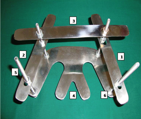
Fig. 1 Occlusal plane analyzer assembled, 1-lateral scale 1, 2-lateral scale 2, 3-front scale, 4-lower stainless steel plate, 5-vertical threaded rods, 6-nuts to fix position of scale with vertical rods.
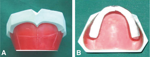
Fig. 2 Adjusted occlusal rim with adapted metal foil on top surface. A: Frontal view, B: Occlusal view.
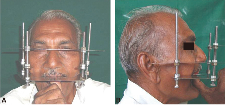
Fig. 3 Establishing plane of occlusion of maxillary rim using occlusal plane analyzer. A: Frontal view, B: Profile view (Using superior point on tragus).
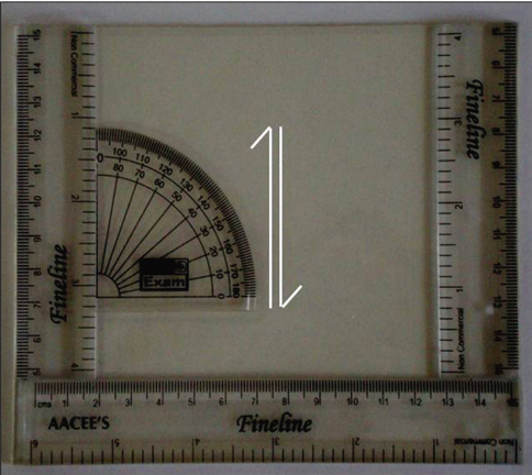
Fig. 4 Angle measuring device with modified protractor.
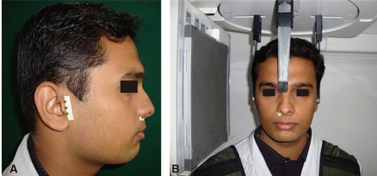
Fig. 5 A: Placement of stainless steel balls at the located points of tragus and ala, B: Adjusting subject with cephalostat for shooting lateral cephalograph.
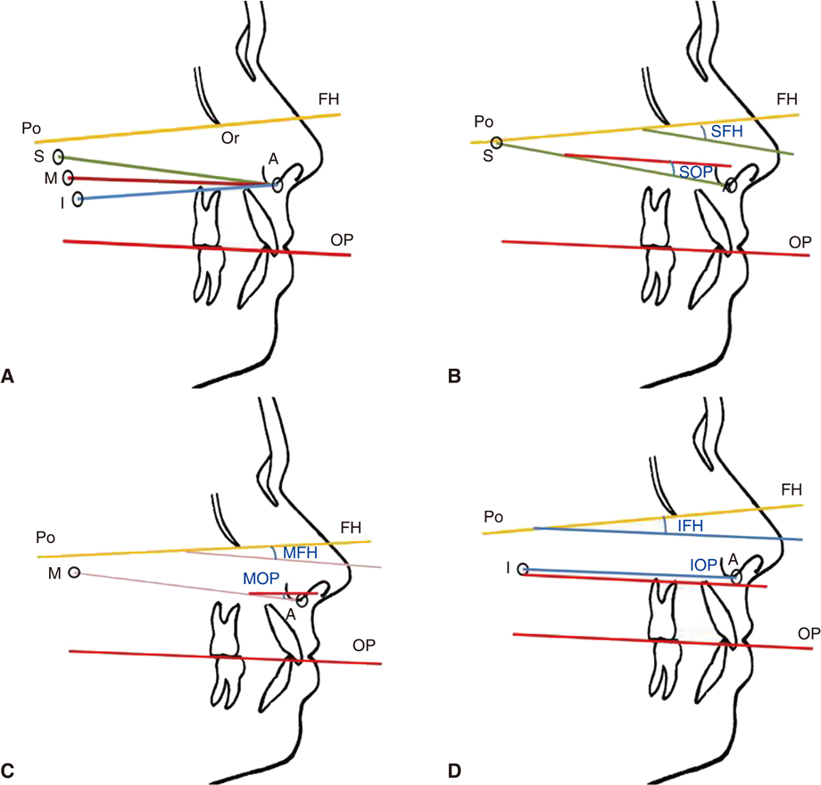
Fig. 6 A: Points and planes used in study, B: Angles measured in study SFH, SOP, C: Angles measured in study MFH, MOP, D: Angles measured in study IFH, IOP.
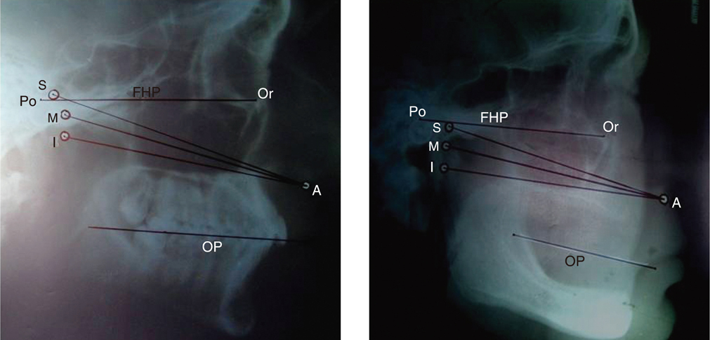
Fig. 7 A: Tracing of lateral cephalograph of dentulous subject, B: Tracing of lateral cephalograph of edentulous subject.
Reference
-
1. Monteith BD. A cephalometric method to determine the angulation of the occlusal plane in edentulous patients. J Prosthet Dent. 1985. 54:81–87.2. Celebić A, Valentić-Peruzović M, Kraljević K, Brkić H. A study of the occlusal plane orientation by intra-oral method (retromolar pad). J Oral Rehabil. 1995. 22:233–236.3. Karkazis HC, Polyzois GL. A study of the occlusal plane orientation in complete denture construction. J Oral Rehabil. 1987. 14:399–404.4. L'Estrange PR, Vig PS. A comparative study of the occlusal plane in dentulous and edentulous subjects. J Prosthet Dent. 1975. 33:495–503.5. Boucher CO, Hickey JC, Zarb GA, Bolender C. Boucher's prosthodontic treatment for edentulous patients. 1985. 9th ed. St. Louis: CV Mosby;243–291.6. Boucher CO. Occlusion in prosthodontics. J Prosthet Dent. 1953. 3:653–656.7. Lundquist DO, Luther WW. Occlusal plane determination. J Prosthet Dent. 1970. 23:489–498.8. Ismail YH, Bowman JF. Position of the occlusal plane in natural and artificial teeth. J Prosthet Dent. 1968. 20:407–411.9. Yasaki M. The height of the occlusion rim and the interocclusal distance. J Prosthet Dent. 1961. 11:26–31.10. Downs WB. Variations in facial relationships; their significance in treatment and prognosis. Am J Orthod. 1948. 34:812–840.11. Athanasiou AE. Orthodontic Cephalometry. 1995. 2nd ed. Philadelphia: Mosby International;255–257.12. Jacobson A. Radiographic Cephalometry. 1992. Chicago: Quintessence Publishing Co;65–75.13. Augsburger RH. Occlusal plane relation to facial type. J Prosthet Dent. 1953. 3:755–770.14. Spratley MH. A simplified technique for determining the occlusal plane in full denture construction. J Oral Rehabil. 1980. 7:31–33.15. van Niekerk FW, Miller VJ, Bibby RE. The ala-tragus line in complete denture prosthodontics. J Prosthet Dent. 1985. 53:67–69.16. Fushima K, Kitamura Y, Mita H, Sato S, Suzuki Y, Kim YH. Significance of the cant of the posterior occlusal plane in class II division 1 malocclusions. Eur J Orthod. 1996. 18:27–40.17. Hartono R. The occlusal plane in relation to facial types. J Prosthet Dent. 1967. 17:549–558.18. Monteith BD. Evaluation of a cephalometric method of occlusal plane orientation for complete dentures. J Prosthet Dent. 1986. 55:64–69.19. Ow RK, Djeng SK, Ho CK. The relationship of upper facial proportions and the plane of occlusion to anatomic reference planes. J Prosthet Dent. 1989. 61:727–733.20. D'Souza NL, Bhargava K. A cephalometric study comparing the occlusal plane in dentulous and edentulous subjects in relation to the maxillomandibular space. J Prosthet Dent. 1996. 75:177–182.21. Jayachandran S, Ramachandran CR, Varghese R. Occlusal plane orientation: a statistical and clinical analysis in different clinical situations. J Prosthodont. 2008. 17:572–575.22. Nairn N. Complete denture prosthetics. 1991. 3rd ed. London: Wright;55–58.23. Nissan J, Barnea E, Zeltzer C, Cardash HS. Relationship between occlusal plane determinants and craniofacial structures. J Oral Rehabil. 2003. 30:587–591.24. Karkazis HC, Polyzois GL. Cephalometrically predicted occlusal plane: implications in removable prosthodontics. J Prosthet Dent. 1991. 65:258–264.25. Solomon EGR, Sridhar Shetty N, Marla V. The morphology of tragus. part II: Reliability of tragus morphology and its reference to established camper's plane. J Inf Proc Syst. 2000. 11:16–22.26. Shigli K, Chetal BR, Jabade J. Validity of soft tissue landmarks in determining the occlusal plane. J Indian Prosthodont Soc. 2005. 5:139–145.
- Full Text Links
-
- Actions
-
Cited
- CITED
-
- Close
- Share
-
- Similar articles
-
- A determination of occlusal plane comparing different levels of the tragus to form ala-tragal line or Camper's line: A photographic study
- Faculty-supervised measurements of the face and of mandibular movements on young adults
- A Case of Cervical Accessory Tragus
- Three Cases of Accessory Tragus
- Reconstruction of Atypical Tragus in Patients with Accessory Tragus or Macrotragus
