Faculty-supervised measurements of the face and of mandibular movements on young adults
- Affiliations
-
- 1Restorative and Prosthetic Dentistry, The Ohio State University College of Dentistry, Columbus, Ohio, USA.
- 2Department of Fixed Prosthodontics, Nihon University School of Dentistry, Tokyo, Japan.
- 3Department of Prosthodontics, Wonkwang University College of Dentistry, Iksan, Korea. dong@wku.ac.kr
- KMID: 2284722
- DOI: http://doi.org/10.4047/jap.2014.6.6.483
Abstract
- PURPOSE
The purpose of this study was to determine the average facial proportions and mandibular movement capacity of 316 first-year dental students who carefully recorded them on each other.
MATERIALS AND METHODS
This early exacting clinical experience was closely supervised by the authors in Columbus, Ohio during 1969-70. Five vertical and six horizontal distances were measured on each subject's face. An ala-tragus line and an occlusal line were drawn on the left side of the face to determine if these two lines were parallel. Measurements of mandibular movements involved maximum normal and hinge opening at the incisors and maximum amounts of right, left lateral and protrusive excursions of the mandible.
RESULTS
The ala width and distance between the tips of upper right and left canine cusps averaged (35.2 mm and 34.8 mm) but with very large individual variations. The distance between ala to occlusal plane lines was 29.9 mm at the tragus and 31.3 mm near the ala. The angle between orbitale and ala-tragus averaged 13.6 degrees.
CONCLUSION
The upper lip length was the most variable and the distance between the pupils was the most stable of the eleven facial measurements. The ala-tragus line and the occlusal plane lines were for all practical purposes parallel. Maximum jaw opening averaged 51.2 mm which was 3.0 times larger than maximal hinge opening of 17.2 mm. The maximum right plus left side jaw excursions (9.2 and 9.4 mm) totaled 18.6 mm, 2.3 times more than the 8.0 mm mean maximum forward protrusion.
Keyword
MeSH Terms
Figure
-

Fig. 1 Landmarks on the face: Trichion (tr) is the point on the hairline in the midline of the forehead, gnathion (gn) is the lowest median landmark on the lower border of the mandible, endocanthion (en) is the point at the inner commissure of the eye fissure, exocanthion (ex) is the outer or lateral eye commisure, (sto) is the imaginary midline point, and cheilion (ch) is the most lateral point on each side of the gently closed lips. Tragions (t) is the notch on the upper margin of the tragus, zygions (zy) is the most lateral point of each zygomatic arch, pupil (p) is the center point of the pupil with the head relaxed and the eyes looking straight forward.
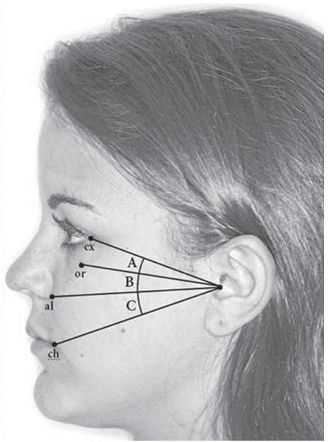
Fig. 2 Depicts the four lines drawn on the side of the faces of 316 dental students in order to measure angles (A, B, and C) below the exocanthion to tragus line with a protractor.
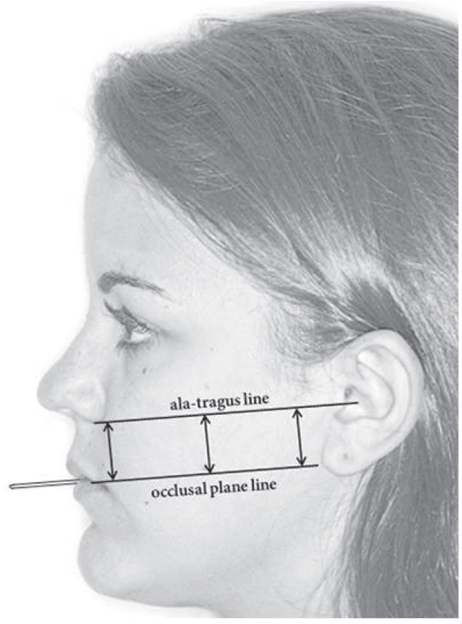
Fig. 3 Two lines were drawn on the left side of each subject's face: one was continuous with and parallel to the tongue depressor which extended about 25 mm in front of the lips while the subject closed firmly on it. The second line was an ala to tragus line. The distance between these two lines was measured in three places: near the ala, at the midpoint, and near the tragus.

Fig. 4 The maximum opening (MO), and hinge opening (HO), was measured between the edges of the upper and lower central incisors using a millimeter ruler with the jaw open. The vertical incisal overlap of the incisors (from dental casts) was added to each mouth measurement in order to include the entire opening distance.

Fig. 5 Measuring the maximum protrusive (MP), maximum left (ML), and maximum right (MR), lateral excursions of the mandible. The distance measured in the mouth between the facial surfaces of the opposing teeth was added to the horizontal overlap of these teeth which had previously been measured on casts in order to include the total amount of jaw excursion in each direction.
Reference
-
1. Peck H, Peck S. A concept of facial esthetics. Angle Orthod. 1970; 40:284–318.2. House MM, Loop JL. Form and color harmony in the dental art. Monograph. Whittier, Calif: MM, House;1939.3. Wehner PJ, Hickey JC, Boucher CO. Selection of artificial teeth. J Prosthet Dent. 1967; 18:222–232.4. Lenner PR. An Introduction to dental anatomy and esthetics. Chicago, IL: Quintessence Publishing Co.;1985. p. 246–271.5. Broadbent TR, Mathews VL. Artistic relationships in surface anatomy of the face: application to reconstructive surgery. Plast Reconstr Surg (1946). 1957; 20:1–17.6. Lee L, Lee KJ. A study of facial proportions and sketching of facial contours. Ear Nose Throat J. 1979; 58:150–158.7. Farkas LG, Katic MJ, Hreczko TA, Deutsch C, Munro IR. Anthropometric proportions in the upper lip-lower lip-chin area of the lower face in young white adults. Am J Orthod. 1984; 86:52–60.8. Farkas LG. Anthropometry of the head and face. 2nd ed. New York: Raven Press;1994.9. Farkas LG, Kolar JC. Anthropometrics and art in the aesthetics of women's faces. Clin Plast Surg. 1987; 14:599–616.10. Ferrario VF, Sforza C, Poggio CE, Serrao G. Facial three-dimensional morphometry. Am J Orthod Dentofacial Orthop. 1996; 109:86–93.11. Sforza C, Grandi G, Binelli M, Dolci C, De Menezes M, Ferrario VF. Age- and sex-related changes in three-dimensional lip morphology. Forensic Sci Int. 2010; 200:182.e1–182.e7.12. Nagle RJ, Sears VH. Denture prosthetics, complete dentures. St. Louis; Toronto, London: CV Mosby Co.;1962. p. 134.13. Lejoyeux J. Prothese complete. Paris: Librairie Maloine S.A.;1967. p. 15.14. Ismail YH, Bowman JF. Position of the occlusal plane in natural and artificial teeth. J Prosthet Dent. 1968; 20:407–411.15. Hickey JC, Zarb GA, Bolender CL. Boucher's prosthodontic treatment for edentulous patients. 9th ed. St. Louis, Toronto, London: CV Mosby Co.;1985. p. 292.16. Gedrange T, Mai R, Richter G, Wolf P, Lupp A, Harzer W. X-ray microanalysis of elements in the masticatory muscle after paresis of the right masseter. J Dent Res. 2005; 84:1026–1030.17. Hützen D, Proff P, Gedrange T, Biffar R, Bernhard O, Kocher T, Kordass B. Occlusal contact patterns-population-based data. Ann Anat. 2007; 189:407–411.18. Posselt U. Movement areas of the mandible. J Prosthet Dent. 1957; 7:375–385.19. Visser A, Naeije M, Hansson TL. The temporal/masseter cocontraction: an electromyographic and clinical evaluation of short-term stabilization splint therapy in myogenous CMD patients. J Oral Rehabil. 1995; 22:387–389.20. Posselt U. Physiology of occlusion and rehabilitation. Oxford: Blackwell;1962. p. 41. p. 56. p. 181.21. Ingervall B. Range of movement of mandible in children. Scand J Dent Res. 1970; 78:311–322.22. Sheppard IM, Sheppard SM. Maximal incisal opening-a diagnostic index? J Dent Med. 1965; 20:13–15.23. Theusner J, Plesh O, Curtis DA, Hutton JE. Axiographic tracings of temporomandibular joint movements. J Prosthet Dent. 1993; 69:209–215.24. Posselt U, Nevstedt P. Registration of the condyle path inclination by intraoral wax records ? its practical value. J Prosthet Dent. 1961; 11:43–47.25. Yatabe M, Zwijnenburg A, Megens CC, Naeije M. The kinematic center: a reference for condylar movements. J Dent Res. 1995; 74:1644–1648.26. Farkas LG, Hreczko TA, Kolar JC, Munro IR. Vertical and horizontal proportions of the face in young adult North American Caucasians: revision of neoclassical canons. Plast Reconstr Surg. 1985; 75:328–338.27. da Vinci L. Trattario della pittura, Bologna. In : Boyd E, editor. Origines of the study of human growth. Portland: University of Oregon Health Science Center Foundation;1980. p. 167.28. Francesca P. Study of the proportion of the head. In : Berrry WA, editor. Drawing the human forms: Methods, Sources, Concepts. NewYork: Van Nostrand Reinhold;1977. p. 116.29. Scheid RC, Weiss G. Woelfel's dental anatomy. 8th ed. Wolters Kluwer Health / Lippincott Williams and Wilkins;2007. p. 263. p. 267. p. 277. p. 288.30. Solberg WK, Woo MW, Houston JB. Prevalence of mandibular dysfunction in young adults. J Am Dent Assoc. 1979; 98:25–34.31. Reicheneder C, Proff P, Baumert U, Gedrange T. Comparison of maximum mouth-opening capacity and condylar path length in adults and children during the growth period. Ann Anat. 2008; 190:344–350.
- Full Text Links
-
- Actions
-
Cited
- CITED
-
- Close
- Share
-
- Similar articles
-
- A study on the morphologic differences between long-face adults and normal-face adults on the lateral and P-A cephalograms
- Differences in opening and protrusive mandibular movements between Class I and II malocclusions in healthy adolescents
- Fabrication of complete dentures using treatment dentures for a patient with erratic mandibular movements: A case report
- The Angles of the Mandible in Korean - Three Dimensional Reconstruction Study
- MANDIBULAR CONTOURING SURGERY BY MULTIPLE STEP SURGICAL CORRECTION WITH ANGLE-SPLITTING OSTECTOMY
