Retrorectal Teratoma with Ovarian Teratoma
- Affiliations
-
- 1Department of Surgery, Ewha Womans University School of Medicine, Seoul, Korea. ralee@ewha.ac.kr
- 2Department of Pathology, Ewha Womans University School of Medicine, Seoul, Korea.
- 3Department of Obstetrics and Gynecology, Ewha Womans University School of Medicine, Seoul, Korea.
- KMID: 2284007
- DOI: http://doi.org/10.12771/emj.2012.35.2.140
Abstract
- No abstract available.
MeSH Terms
Figure
-

Fig. 1 Pelvic ultrasonography findings. (A) 7.3×6.2 cm sized left ovary with inhomogeneous cystic lesion is noted. (B) Normal sized right ovary is demonstrated.
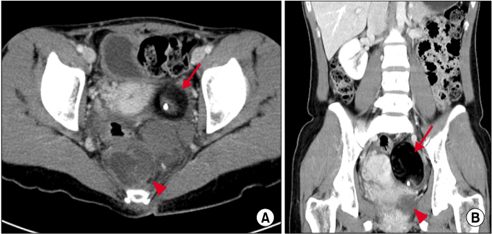
Fig. 2 Abdomen & pelvis computed tomography findings. CT images show 5.2×5.8 cm sized cystic mass (indicated by arrow) in the left ovary and 5.5×6.3 cm sized mass (indicated by arrowhead) composed of calcification, fat and soft tissue adjacent to rectum in the presacral area. (A) Axial view. (B) Coronal view.

Fig. 3 Microscopic findings. Histological findings of the resected specimen in left ovarian mass (A) and retroporitoneal mass (B) are composed of keratinizing stratified squamous epithelium associated with the hair and mucus, which are compatible with mature cystic teratoma (H&E stain, ×40).
Reference
-
1. Terada T. Mature teratoma of the rectum. Gastrointest Endosc. 2010. 71:1068–1069.2. Kaeser MA, McDonald JK, Kettner NW. A calcific pelvic mass in a woman with chronic spinal pain: a case of mature cystic teratoma. J Chiropr Med. 2011. 10:327–332.3. Baek EH, Kim KI, Lee AR, Gang G, Kwon YS. Periappendiceal mature cystic teratoma successfully treated with laparoendoscopic surgery. Am Surg. 2012. 78:70–71.4. Moawad NS, Starks D, Ashby K. Ectopic ovarian teratoma of the uterosacral ligament associated with a large ovarian dermoid. J Minim Invasive Gynecol. 2008. 15:523–524.5. Khoo CK, Chua I, Siow A, Chern B. Parasitic dermoid cyst of the pouch of Douglas: a case report. J Minim Invasive Gynecol. 2008. 15:761–763.6. Kahraman K, Kurtay G, Kiremitci S. A presacral dermoid cyst with extremely high serum CA19-9 level. J Obstet Gynaecol. 2012. 32:103–104.7. La Quaglia MP, Feins N, Eraklis A, Hendren WH. Rectal duplications. J Pediatr Surg. 1990. 25:980–984.8. Nishie A, Yoshimitsu K, Honda H, Irie H, Aibe H, Shinozaki K, et al. Presacral dermoid cyst with scanty fat component: usefulness of chemical shift and diffusion-weighted MR imaging. Comput Med Imaging Graph. 2003. 27:293–296.9. Dahan H, Arrivé L, Wendum D, Docou le Pointe H, Djouhri H, Tubiana JM. Retrorectal developmental cysts in adults: clinical and radiologic-histopathologic review, differential diagnosis, and treatment. Radiographics. 2001. 21:575–584.10. Guillem P, Ernst O, Herjean M, Triboulet JP. Retrorectal tumors: an assessment of the abdominal approach. Ann Chir. 2001. 126:138–142.
