Korean J Phys Anthropol.
2014 Sep;27(3):165-171. 10.11637/kjpa.2014.27.3.165.
Morphometric Analysis of the Skull by Moire Contourography
- Affiliations
-
- 1Department of Anatomy, Soonchunhyang University, Cheonan, Korea. mdeornfl@sch.ac.kr
- KMID: 2283733
- DOI: http://doi.org/10.11637/kjpa.2014.27.3.165
Abstract
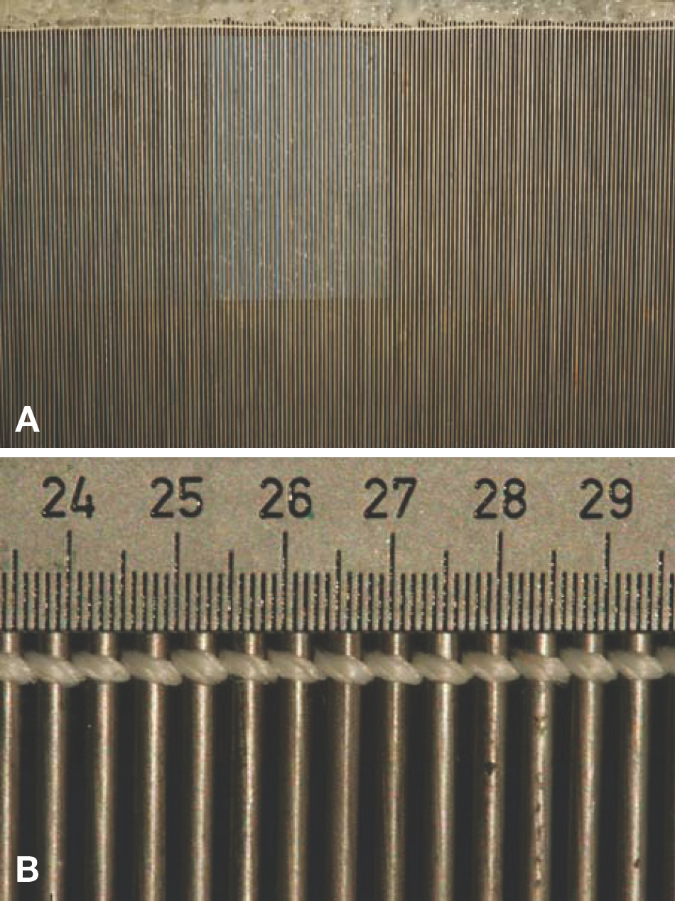
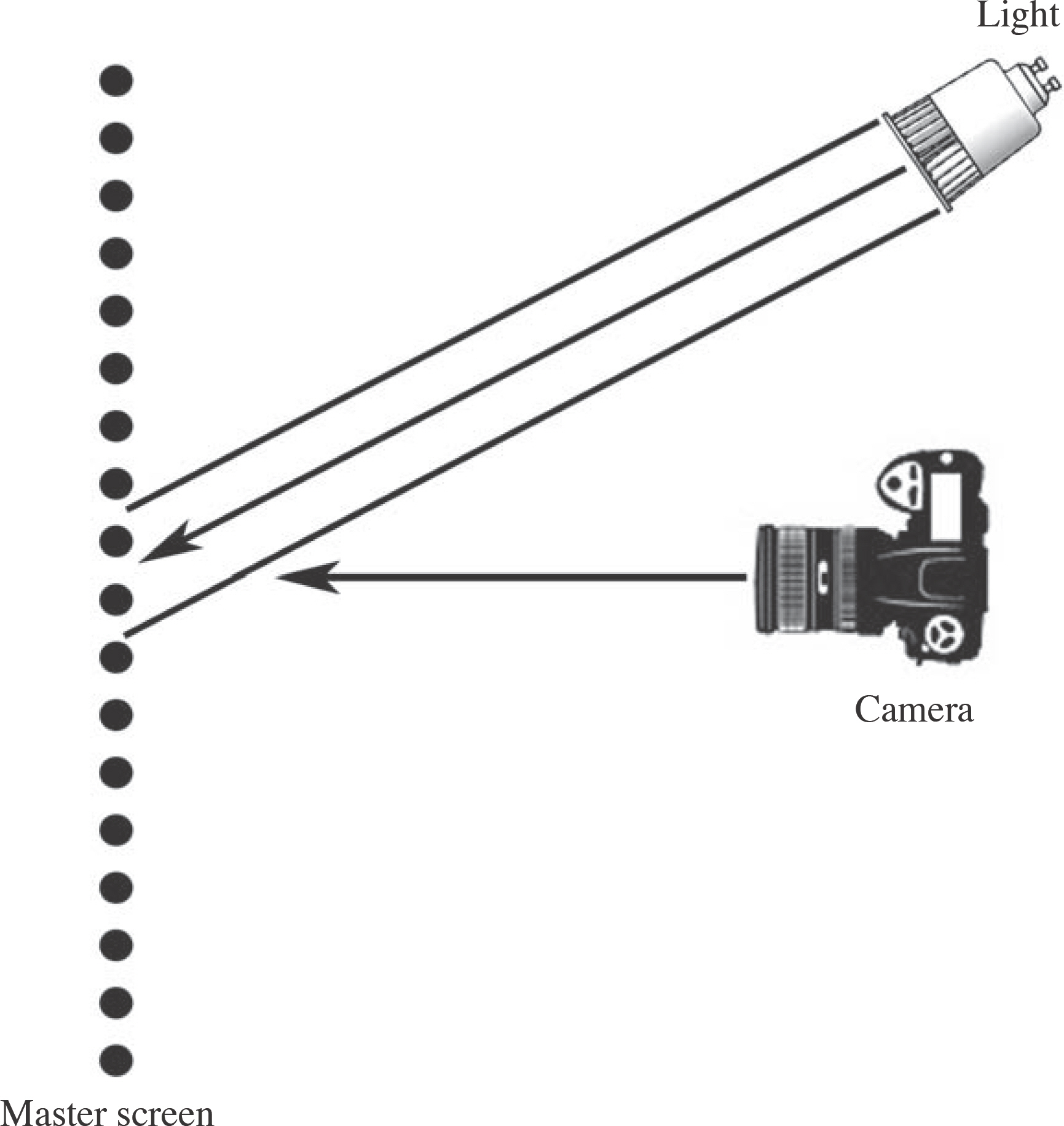
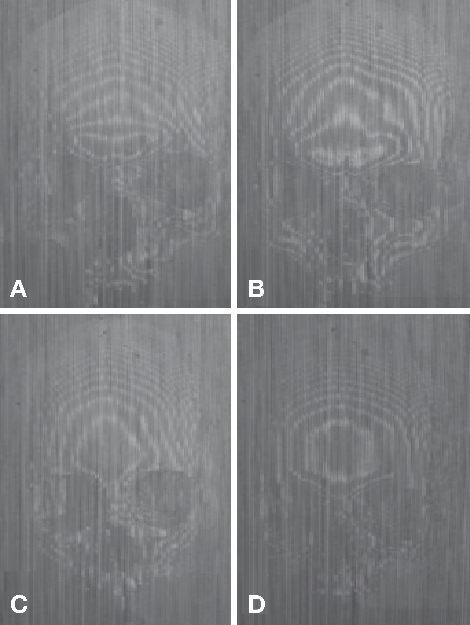
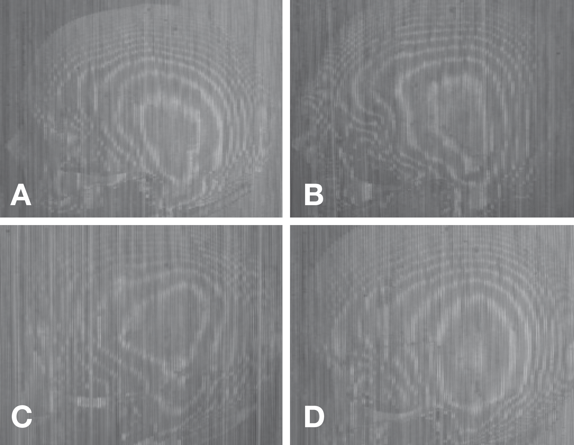
- The non-metric analysis of the skulls is very useful for estimating sex and determination of ancestry, the accuracy tends to depend on the amount of experiences of the observers, and so inter-observer errors might be happened. Many researchers are trying to find out more objective methods for determination of ancestry. The purpose of this presentation is to show the usefulness of moire contourography for analyzing the skull. The master screen that is similar to the gratings was made by steel rods, which were arranged as equally spaced parallel lines. Halogen light source was illuminated by lantern slide projector. The skeletal materials were documented crania, composed of 87 male and 47 female, from William M. Bass Donated Skeletal Collection housed at the Department of Anthropology, University of Tennessee. The skulls were placed just behind the master screen as anatomical position using cubic craniophore. The angle between the light source and camera was 65degrees, the distance between camera and the master screen was 1.2 m. Frontal view, left lateral and right lateral view were taken. From the frontal view, fringe patterns were analyzed for first five contour lines which were mainly located around the Glabella. The results were as followed; Type I for male was 53% and female was 4%; Type II for male was 29% and female was 2%; Type III for male was 2% and female was 15%; Type IV for male was 6% and female was 55%. From the lateral view, fringe patterns were analyzed for first four contour lines. However, first and second contour lines were critical to determine the shape and the results were as followed; Type I for male was 52% and female was 22%; Type II for male was 38% and female was 26%; Type III for male was 8% and female was 17%; Type IV for male was 2% and female was 35%. According to this study, different fringe patterns might be dependent on the degree of development of bone marker such as Glabella, Supercillary arch, Euryon and Mastoid process. For example, Supercillary arches were very well developed and slope of forehead above the Glabella was declined, fringe pattern showed reverse triangle shape. If Supercillary arches were poorly developed and slope of forehead above the Glabella was flat, fringe pattern showed home plate shape. The present research shows that moire contourography might be used as more objective methods for estimating sex. And it would be helpful to determine the ancestry when the lateral aspects were analyzed. In the future, continuing study need to be performed with same master screen for different ancestry.
Keyword
Figure
Reference
-
References
1. Martin R. Lehrbuch der Anthropologie, Zeiter Band: Kran-iologie, Osteologie. Vol. 1:2nd ed.Jena, Germany: Verlag Gustav Fischer;1928.2. Krogman WM, can MY. The human skeleton in forensic medicine. 1st ed.Springfield, Illinois: Charles C Thomas Publisher;1986.3. Hefner JT. Cranial nonmetric variation and estimation ancestry. J Forensic Sci. 2009; 54:985–95.4. Stewart TD. Essentials of forensic anthropology; Especially as developed in the United State. 1st ed.Springfield, Illinois: Charles C Thomas Publisher;1979. p. 85–127.5. White TD, Folkens PA. The human bone manual. 1st ed.San Diego, California: Elsevier Academic Press;2005. p. 8–9. 385–98.6. Takasaki H. Moiré topography. Appl Optics. 1970; 9:1457–72.
Article7. Kanazawa E, Terada H, Masumi S. An application of moiré contourography for the evaluation of surgical operation on patients with spinal scoliosis. Kitasato Med. 1975; 5:75–81.8. Otsuka Y, Shinoto A, Inoue S. Mass school screening for early detection of scoliosis by use moiré topography camera and low dose x-ray imaging. Rinsho Seikei Geka. 1979; 14:973–84.9. Otsuka Y. Moiré topography. Its application to vertebral deformity diagnostics. Shoni Igaku. 1980; 13:1086–110.10. Moreland MS, Pope MH, Armstrong GWD. Moiré Fringe Topography and Spinal Deformity. New York: Pergamon Press;1981.11. Han SH, Kim IB, Kim YH, Park DK, Kim DW. Anthropological analysis of the Korean skull by Moiré contourography. Korean J Phy Anthropol. 1998; 11:223–36. Korean.12. Park DK, Ra JJ, Park KH, Ko JS, Kim DI, Kim YS, et al. Determination of sex in Korean using atlas. Korean J Phy Anthropol. 2009; 22:205–12. Korean.13. Forensic Anthropology Center. WM Bass Donated Skeletal Collection [Internet]. Knoxville, TN. Available from:. http://fac.utk.edu/collection.html.14. Creath K, Schmit J, Wyant JC. Optical Metrology of diffuse surfaces. Malacara D, editor. Optical shop testing. New Jersy: John Wiley & Sons;2007. p. 756–83.
Article15. Bass WM. Human osteology; a laboratory and field manual.5th ed. Springfield, MO: Missouri Archaeological Society;2009.16. Hefner JT, Emanovsky PD, Byrd J, Ousley SD. The value of experience, education, and methods in ancestry prediction. Proceedings of the 59th Annual Meeting of the American Academy of Forensic Sciences, 2007 Feb 19–24; San Antonio, Colorado Srpings: American Academy of Forensic Sciences. 2007.
- Full Text Links
- Actions
-
Cited
- CITED
-
- Close
- Share
- Similar articles
-
- Anthropological Analysis of the Korean Skulls by Moire Contourography
- Upper Face Characteristics According to the Sa -sang Constitution
- A Study on the Korean Orbital and Cranial Index Using 3D Skull CT Image and Morphometric Analysis
- Evaluation of morphometric features of fossa navicularis using cone-beam computed tomography in a Turkish subpopulation
- Adolescent Scoliosis Screening in Nara City Schools: A 23-Year Retrospective Cross-Sectional Study


