Voxel-Wise Analysis of Diffusion Tensor Imaging for Clinical Outcome of Cochlear Implantation: Retrospective Study
- Affiliations
-
- 1Department of Molecular Medicine, Kyungpook National University School of Medicine, Daegu, Korea.
- 2Department of Radiology, Kyungpook National University School of Medicine, Daegu, Korea.
- 3Department of Medical & Biological Engineering, Kyungpook National University School of Medicine, Daegu, Korea.
- 4Department of Otorhinolaryngology, Kyungpook National University School of Medicine, Daegu, Korea. leeshu@knu.ac.kr
Abstract
OBJECTIVES
To evaluate retrospectively, the possible difference in diffusion tensor imaging (DTI) metric of fractional anisotropy (FA) between good and poor surgical outcome cochlear implantation (CI) patients using investigator-independent voxel-wise analysis.
METHODS
Eighteen patients (11 males, 7 females; mean age, 5.9 years) with profound sensorineural hearing loss underwent DTI scans using a 3.0 Tesla magnetic resonance scanner. Among the 18 patients, 10 patients with categories of auditory performance (CAP) score over 6 were classified into the good outcome group and 8 patients with CAP score below 6 were classified into the poor outcome group. The diffusion tensor scalar measure was calculated from the eigenvalues of the tensor on a voxel-by-voxel basis from each subject and two-sample t-test evaluation between good and poor outcome subjects were performed for each voxel of FA values, across the entire brain, with a voxel-wise intensity threshold of P<0.0005 (uncorrected) and a contiguous cluster size of 64 voxels. Individual values of FA were measured by using the region-of-interest based analysis for correlation analysis with CAP scores, open sentence and open word scores.
RESULTS
Two-sample t-test evaluation using SPM voxel-wise analysis found significantly higher FA values at the several brain areas including Broca's area, genu of the corpus callosum, and auditory tract in good outcome subjects compared to poor outcome subjects. Correlation analyses between FA and CAP scores, open sentence and open word scores revealed strong correlations at medial geniculate nucleus, Broca's area, genu of the corpus callosum and auditory tract.
CONCLUSION
Investigator-independent voxel-based analysis of DTI image demonstrated that good outcome subjects showed better neural integrity at brain areas associated with language and auditory functions, suggesting that the conservation of microstructural integrity of these brain areas is important. Preoperative functional imaging may be helpful for CI.
MeSH Terms
Figure
-
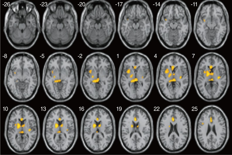
Fig. 1 Two sample statistical parametric mapping (SPM) axial maps rendered on to a normalized T1-weighted MR image, shows areas of significant decrease in fractional anisotropy (FA) values in poor outcome subjects compared to good outcome subjects (thresholded at uncorrected P<0.0005 and the extent of 16 clusters).

Fig. 2 The correlations between fractional anisotropy (FA) values and categories of auditory performance (CAP) scores at the selected brain regions shown in Fig. 1. The lines are the result of a linear regression.

Fig. 3 The correlations between fractional anisotropy (FA) values and open-set sentence perception scores at the selected brain regions shown in Fig. 1. The lines are the result of a linear regression.
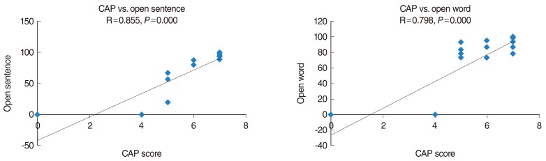
Fig. 4 The correlations between categories of auditory performance (CAP) scores and open-set sentence, open-set word perception scores. The lines are the result of a linear regression.
Reference
-
1. Moore DR, Shannon RV. Beyond cochlear implants: awakening the deafened brain. Nat Neurosci. 2009; 6. 12(6):686–691. PMID: 19471266.
Article2. Svirsky MA, Teoh SW, Neuburger H. Development of language and speech perception in congenitally, profoundly deaf children as a function of age at cochlear implantation. Audiol Neurootol. 2004; Jul-Aug. 9(4):224–233. PMID: 15205550.
Article3. Basser PJ, Pierpaoli C. A simplified method to measure the diffusion tensor from seven MR images. Magn Reson Med. 1998; 6. 39(6):928–934. PMID: 9621916.4. Beaulieu C. The basis of anisotropic water diffusion in the nervous system: a technical review. NMR Biomed. 2002; Nov-Dec. 15(7-8):435–455. PMID: 12489094.5. Hasan KM, Gupta RK, Santos RM, Wolinsky JS, Narayana PA. Diffusion tensor fractional anisotropy of the normal-appearing seven segments of the corpus callosum in healthy adults and relapsing-remitting multiple sclerosis patients. J Magn Reson Imaging. 2005; 6. 21(6):735–743. PMID: 15906348.
Article6. Chang Y, Lee SH, Lee YJ, Hwang MJ, Bae SJ, Kim MN, et al. Auditory neural pathway evaluation on sensorineural hearing loss using diffusion tensor imaging. Neuroreport. 2004; 8. 06. 15(11):1699–1703. PMID: 15257130.
Article7. Bhagat YA, Beaulieu C. Diffusion anisotropy in subcortical white matter and cortical gray matter: changes with aging and the role of CSF-suppression. J Magn Reson Imaging. 2004; 8. 20(2):216–227. PMID: 15269946.
Article8. Silk TJ, Vance A, Rinehart N, Bradshaw JL, Cunnington R. White-matter abnormalities in attention deficit hyperactivity disorder: a diffusion tensor imaging study. Hum Brain Mapp. 2009; 9. 30(9):2757–2765. PMID: 19107752.
Article9. Scherfler C, Schocke MF, Seppi K, Esterhammer R, Brenneis C, Jaschke W, et al. Voxel-wise analysis of diffusion weighted imaging reveals disruption of the olfactory tract in Parkinson's disease. Brain. 2006; 2. 129(Pt 2):538–542. PMID: 16272163.
Article10. Kim Y, Jeong KS, Song HJ, Lee JJ, Seo JH, Kim GC, et al. Altered white matter microstructural integrity revealed by voxel-wise analysis of diffusion tensor imaging in welders with manganese exposure. Neurotoxicology. 2011; 1. 32(1):100–109. PMID: 21111757.
Article11. Watkins KE, Paus T, Lerch JP, Zijdenbos A, Collins DL, Neelin P, et al. Structural asymmetries in the human brain: a voxel-based statistical analysis of 142 MRI scans. Cereb Cortex. 2001; 9. 11(9):868–877. PMID: 11532891.
Article12. Eckert MA, Leonard CM, Wilke M, Eckert M, Richards T, Richards A, et al. Anatomical signatures of dyslexia in children: unique information from manual and voxel based morphometry brain measures. Cortex. 2005; 6. 41(3):304–315. PMID: 15871596.
Article13. Emmorey K, Allen JS, Bruss J, Schenker N, Damasio H. A morphometric analysis of auditory brain regions in congenitally deaf adults. Proc Natl Acad Sci U S A. 2003; 8. 100(17):10049–10054. PMID: 12904582.
Article14. Karas GB, Burton EJ, Rombouts SA, van Schijndel RA, O'Brien JT, Scheltens P, et al. A comprehensive study of gray matter loss in patients with Alzheimer's disease using optimized voxel-based morphometry. Neuroimage. 2003; 4. 18(4):895–907. PMID: 12725765.
Article15. Le Bihan D, Mangin JF, Poupon C, Clark CA, Pappata S, Molko N, et al. Diffusion tensor imaging: concepts and applications. J Magn Reson Imaging. 2001; 4. 13(4):534–546. PMID: 11276097.
Article16. Ono J, Harada K, Takahashi M, Maeda M, Ikenaka K, Sakurai K, et al. Differentiation between dysmyelination and demyelination using magnetic resonance diffusional anisotropy. Brain Res. 1995; 2. 671(1):141–148. PMID: 7728526.
Article17. Rai V, Nath K, Saraswat VA, Purwar A, Rathore RK, Gupta RK. Measurement of cytotoxic and interstitial components of cerebral edema in acute hepatic failure by diffusion tensor imaging. J Magn Reson Imaging. 2008; 8. 28(2):334–341. PMID: 18626948.
Article18. Shibata DK. Differences in brain structure in deaf persons on MR imaging studied with voxel-based morphometry. AJNR Am J Neuroradiol. 2007; 2. 28(2):243–249. PMID: 17296987.19. Bookstein FL. "Voxel-based morphometry" should not be used with imperfectly registered images. Neuroimage. 2001; 12. 14(6):1454–1462. PMID: 11707101.
- Full Text Links
-
- Actions
-
Cited
- CITED
-
- Close
- Share
-
- Similar articles
-
- Inter-Vendor and Inter-Session Reliability of Diffusion Tensor Imaging: Implications for Multicenter Clinical Imaging Studies
- Principle and Experiments in Diffusion Tensor Imaging
- Diffusion Tensor Imaging: Exploring the Motor Networks and Clinical Applications
- Cochlear Implant Failure due to Cochlear Nerve Deficiency in a Child with Normal Internal Auditory Canal
- Multi-slice Multi-echo Pulsed-gradient Spin-echo (MePGSE) Sequence for Diffusion Tensor Imaging MRI: A Preliminary Result
